. 2019 Dec;30(12):2293-2305.
doi: 10.1681/ASN.2019020148. Epub 2019 Sep 23.

Kidney Regeneration in Later-Stage Mouse Embryos via Transplanted Renal Progenitor Cells

Affiliations

Kidney Regeneration in Later-Stage Mouse Embryos via Transplanted Renal Progenitor Cells

Shuichiro Yamanaka et al. J Am Soc Nephrol. 2019 Dec.

Abstract

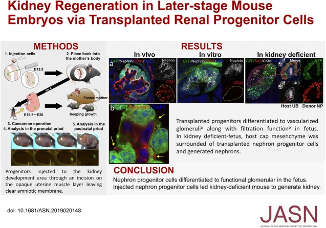
Background: The limited availability of donor kidneys for transplantation has spurred interest in investigating alternative strategies, such as regenerating organs from stem cells transplanted into animal embryos. However, there is no known method for transplanting cells into later-stage embryos, which may be the most suitable host stages for organogenesis, particularly into regions useful for kidney regeneration.

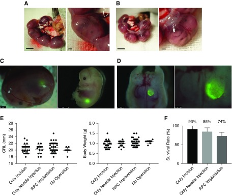
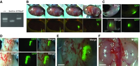
Methods: We demonstrated accurate transplantation of renal progenitor cells expressing green fluorescent protein to the fetal kidney development area by incising the opaque uterine muscle layer but not the transparent amniotic membrane. We allowed renal progenitor cell-transplanted fetuses to develop for 6 days postoperatively before removal for analysis. We also transplanted renal progenitor cells into conditional kidney-deficient mouse embryos. We determined growth and differentiation of transplanted cells in all cases.

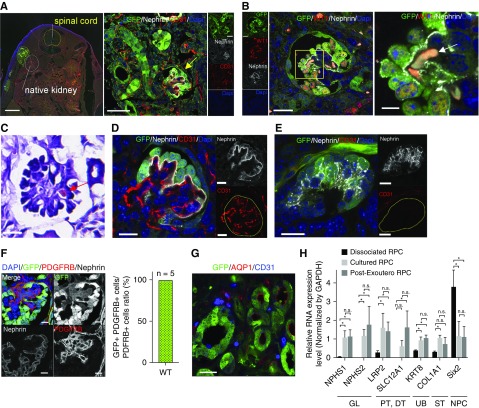
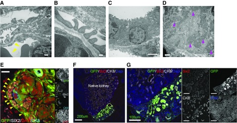
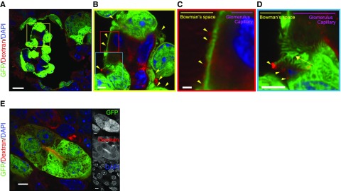
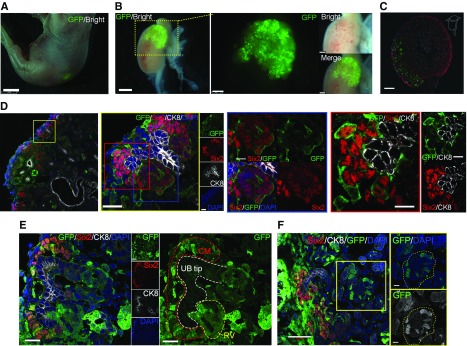
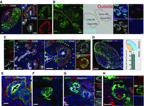
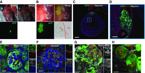
Results: Renal progenitor cell transplantation into the retroperitoneal cavity of fetuses at E13-E14 produced transplant-derived, vascularized glomeruli with filtration function and did not affect fetal growth or survival. Cells transplanted to the nephrogenic zone produced a chimera in the cap mesenchyme of donor and host nephron progenitor cells. Renal progenitor cells transplanted to conditional kidney-deficient fetuses induced the formation of a new nephron in the fetus that is connected to the host ureteric bud.

Conclusions: We developed a cell transplantation method for midstage to late-stage fetuses. In vivo kidney regeneration from renal progenitor cells using the renal developmental environment of the fetus shows promise. Our findings suggest that fetal transplantation methods may contribute to organ regeneration and developmental research.

Keywords: kidney development; kidney regeneration; nephron; pluripotent stem cell; progenitor; stem cell.


Figures

None
Graphical abstract
Figure 1.
Figure 1.
Transplantation of renal progenitor cells into the fetus allows surviving without inhibiting growth. (A) Photomicrographs of a fetus in utero at E13.5. (B) Fetus in utero after cutting the uterine muscle layer. (C and D) Visualization of GFP-expressing transplanted RPCs in the retroperitoneal cavity at E13.5. Images demonstrate a fetus removed after cell injection and dissected for validation of cell transplantation. (E, left) Crown-rump length (CRL) and (right) body weight at E19.5 were compared across the RPC-injected experimental group (n=35), a noninjected (puncture only) group (n=15), and a sham-operated group (n=20). (F) The survival rate was calculated as a percentage of the number of surviving fetuses 6 days after the operation (sham operation, n=14; puncture only, n=20; and RPC injection, n=19). There was no significant difference between groups. Bars represent means±SEM. P<0.05, Mann–Whitney U test. Scale bars, 1 mm in (A), (B), and left panel of (C) and (D); 2 mm in right panel of (C); 250 µm in right panel of (D).
Figure 2.
Figure 2.
Analysis of nephrons derived from RPC regenerated inside the retroperitoneal cavity of fetus. (A) In immunofluorescence imaging, transplanted RPCs expressing GFP were present outside the host kidney in the retroperitoneum. Mature glomeruli with capillary loops (yellow arrow) derived from transplanted cells can be seen. (B and C) Erythrocytes (white arrow in [B] and red arrow in [C]) and blood flow were identified. (C) Hematoxylin and eosin stain. (D) In vivo glomeruli had capillary loops containing host embryo vascular endothelial cells (CD31+ and GFP). (E) In vitro glomeruli without vascular endothelial cells (CD31+). (F) PDGFRb-positive cells in GFP-expressing glomeruli. All PDGFRb-positive cells expressed GFP. Refer to Supplemental Methods. (G) Transplanted cells expressing tubule markers AQP1 (red). (H) Quantified PCR analysis of nephron genes. Kruskal–Wallis test was for comparison. Error bars indicate SEM; *P<0.05; n=6. Scale bars, 500 µm in the left panel of (A); 50 µm in the right panel of (A); 20 µm in left panel of (B) and (C); 5 µm in right panel of (B); 20 µm in (D), (E), and (G); 10 µm in (F). DAPI, 4′,6-diamidino-2-phenylindole; DT, distal tubule; GAPDH, glyceraldehyde-3-phosphate dehydrogenase; GL, glomerulus; PT, proximal tubule; ST, stromal tissue; WT, wild type.
Figure 3.
Figure 3.
Nephrons regenerate inside the retroperitoneal cavity are highly differentiated but do not integrate with the host kidney beyond the renal capsule. (A and B) Podocytes and slit diaphragms in glomeruli derived from transplanted cells. Erythrocytes indicated by yellow triangles on the endothelial side. (C) Brush border in the lumen of the proximal tubule. (D) There were abundant mitochondria in the cells (magenta triangles). (E) GFP-positive cells surrounded Six2-positive NPCs and Six2-negative cells (yellow triangle) in the cap mesenchyme. (F and G) Donor UB cells differentiated into UB tip and collecting duct but did not integrate with the host UB. Scale bars, 500 nm in (A) and (D); 100 nm in (B); 2 µm in (C); 20 µm in (E); 200 µm in (F); 100 µm in (G). CK8, Cytokeratin 8; DAPI, 4′,6-diamidino-2-phenylindole.
Figure 4.
Figure 4.
Generated glomeruli displayed podocytes and filtration function. (A–C) Fluorescence-labeled dextrans in blood were filtered to the Bowman’s space side in the glomerulus (displayed filtered dextran with yellow triangles) derived from RPCs implanted in the wild-type mice. (D) Podocytes formed a filtration slit (yellow arrow). (E) Dextran was seen in the uriniferous tubule cavity. Scale bars, 20 µm in (A); 2 µm in (B) and (D); 1 µm in (C); 5 µm in (E). DAPI, 4′,6-diamidino-2-phenylindole.
Figure 5.
Figure 5.
Transplantation of RPC under the renal capsule enables integration with the host kidney but is a chimeric structure. (A) Fetus at E19.5 with RPCs expressing GFP (day 6 postinjection). (B) GFP-expressing progenitor cells integrated into the host kidney. (C and D) Integration of exogenous NPCs into host cap mesenchyme. Higher magnification of (D) left panel (blue frame panel) showing transplanted NPCs in the host renal vesicle (RV), and (red frame panel) chimerism of NPCs in cap mesenchyme (CM) visualized by GFP. (E) Mosaicism in the RV. (F) Chimeric glomeruli-like mosaics contributed by the host and donor NPCs. The yellow dotted line indicates the glomerulus. Scale bars, 2 mm in (A); 1 mm in left panel of (B); 250 µm in right panel of (B); 200 µm in (C); 20 µm in left panel and yellow-framed panel of (D) and in (E); 10 µm in blue- and red-framed panel of (D); 50 µm in (F). CK8, Cytokeratin 8; DAPI, 4′,6-diamidino-2-phenylindole.
Figure 6.
Figure 6.
Transplanted RPC form kidney in the renal-deficient fetus. (A) Genotyping of the fetus (Six2-GFPCre+; iDTR+). (B) GFP-expressing RPCs transplanted into the renal development area of the kidney-deficient embryo (Six2-GFPCre+; iDTR+) at E13.5. (C) Fetuses 6 days after transplantation. (D and E) Fetal kidney 6 days after transplantation. Note the unilateral GFP expression (right side). (F) Kidney of wild-type B6 mouse at E19.5. a, adrenal gland; k, fetal kidney; u, fetal ureter.
Figure 7.
Figure 7.
RPC transplanted in a renal-deficient fetus connect with the host ureteric bud to form nephrons. (A) Immunolabeling of transplanted cells integrated into the host kidney. Transplanted NPCs gathered around the UB tip and formed cap mesenchyme (CM). (B) Transplanted cells present outside the kidney self-organized to form CM with GFP-expressing UB and NPCs (outside). The transplanted cells that reached the host nephrogenic zone aggregated around the host UB tip (GFP negative) and formed new CM (inside). (C) GFP-negative cells (yellow triangle) surrounded Six2-positive NPCs in the CM. (D) Overall, 33% of the regenerated cap mesenchyme was integrated with the host UB tip (counted to a total of 119 tips, n=4 slices). Refer to Supplemental Methods. (E–H) Transplanted cells expressing tubule and nephron markers. (G and H) Mature glomeruli with capillary loops derived from transplanted GFP cells. (H) AQP1 is expressed in structures associated with the developing renal vasculature. The expression of AQP1 was observed inside regenerated glomeruli. It was noted as a host-derived tissue because it was GFP negative. Scale bars, 100 µm in (A) and (B); 50 µm in (C); 10 µm in (E) and (F); 20 µm in (G) and (H). CK8, Cytokeratin 8; DAPI, 4′,6-diamidino-2-phenylindole.
Figure 8.
Figure 8.
Regenerating nephrons inside the retroperitoneal cavity of the Wild-type mouse are able to be maintained after birth. (A and B) Transplanted GFP-RPCs adhered to the native kidney and were connected to the host’s blood vessels. (C) Kidney was collected and immunofluorescence staining was performed. (D) Transplanted GFP-RPCs partially penetrated into the fetal kidney at postnatal day 14 (P14). (E) The transplanted GFP-RPCs differentiated into nephrons with expressed nephrin, and CD31-positive endothelial cells were observed in the glomerulus under high magnification (63× magnification). (F) As control, a normal B6 fetal glomerulus on P14. (G) The transplanted GFP-RPC expressed a proximal tubular marker (Megalin). A site positive for CDH1 (collecting duct and distal tubule marker) and negative for CK8 (collecting duct marker) indicates a distal tubule. (H) Proximal tubule in the high magnification. Scale bars, 2 mm in (A); 250 µm in (B); 500 µm in (C); 20 µm in (D) and (G); 10 µm in (E), (F), and (H). Ao, Aorta; CDH1, Cadherin 1; CK8, Cytokeratin 8; DAPI, 4′,6-diamidino-2-phenylindole.

Comment in

References

    1. Liyanage T, Ninomiya T, Jha V, Neal B, Patrice HM, Okpechi I, et al. .: Worldwide access to treatment for end-stage kidney disease: A systematic review. Lancet 385: 1975–1982, 2015 - PubMed
    1. Yokoo T, Ohashi T, Shen JS, Sakurai K, Miyazaki Y, Utsunomiya Y, et al. .: Human mesenchymal stem cells in rodent whole-embryo culture are reprogrammed to contribute to kidney tissues. Proc Natl Acad Sci U S A 102: 3296–3300, 2005 - PMC - PubMed
    1. Yokoo T, Fukui A, Ohashi T, Miyazaki Y, Utsunomiya Y, Kawamura T, et al. .: Xenobiotic kidney organogenesis from human mesenchymal stem cells using a growing rodent embryo. J Am Soc Nephrol 17: 1026–1034, 2006 - PubMed
    1. Wu J, Platero-Luengo A, Sakurai M, Sugawara A, Gil MA, Yamauchi T, et al. : Interspecies chimerism with mammalian pluripotent stem cells. Cell 168: 473–486.e15, 2017 - PMC - PubMed
    1. Wu J, Greely HT, Jaenisch R, Nakauchi H, Rossant J, Belmonte JC: Stem cells and interspecies chimaeras. Nature 540: 51–59, 2016 - PubMed

Publication types