. 2019 Oct 8;116(41):20545-20555.
doi: 10.1073/pnas.1906663116. Epub 2019 Sep 23.

Transcriptional control of lung alveolar type 1 cell development and maintenance by NK homeobox 2-1

Affiliations

Transcriptional control of lung alveolar type 1 cell development and maintenance by NK homeobox 2-1

Danielle R Little et al. Proc Natl Acad Sci U S A. .

Abstract

The extraordinarily thin alveolar type 1 (AT1) cell constitutes nearly the entire gas exchange surface and allows passive diffusion of oxygen into the blood stream. Despite such an essential role, the transcriptional network controlling AT1 cells remains unclear. Using cell-specific knockout mouse models, genomic profiling, and 3D imaging, we found that NK homeobox 2-1 (Nkx2-1) is expressed in AT1 cells and is required for the development and maintenance of AT1 cells. Without Nkx2-1, developing AT1 cells lose 3 defining features-molecular markers, expansive morphology, and cellular quiescence-leading to alveolar simplification and lethality. NKX2-1 is also cell-autonomously required for the same 3 defining features in mature AT1 cells. Intriguingly, Nkx2-1 mutant AT1 cells activate gastrointestinal (GI) genes and form dense microvilli-like structures apically. Single-cell RNA-seq supports a linear transformation of Nkx2-1 mutant AT1 cells toward a GI fate. Whole lung ChIP-seq shows NKX2-1 binding to 68% of genes that are down-regulated upon Nkx2-1 deletion, including 93% of known AT1 genes, but near-background binding to up-regulated genes. Our results place NKX2-1 at the top of the AT1 cell transcriptional hierarchy and demonstrate remarkable plasticity of an otherwise terminally differentiated cell type.

Keywords: NK homeobox 2-1; alveolar type 1 cell; alveologenesis; lung development; transcriptional control.


Conflict of interest statement

The authors declare no conflict of interest.

Figures

Fig. 1.
Fig. 1.
NKX2-1 is required in AT1 cells for alveologenesis. (A) Schematics of sample preparation to obtain an en face view of complete AT1 cells (traced from an immunostained P3 lung), whose conventional sectional view captures part of a cell as a line bordering the airspace. (B) Confocal en face view of immunostained RosaSun1GFP/+; ShhCre/+ lung strips showing that NKX2-1 is expressed by all epithelial cells, whose nuclei are genetically marked with GFP, including all AT1 cells as identified by the expanded cell size outlined by ECAD and absence of LAMP3 (arrowheads). At least 2 mice were examined at each time point with consistent results. (C) Confocal en face view of immunostained littermate lung strips showing specific loss of NKX2-1 in GFP-marked, expanded AT1 cells in the mutant (open versus filled arrowheads). At least 3 lungs per genotype were examined with consistent results. (D) Left 3 columns: H&E stained sections from littermate lungs. At least 3 lungs per genotype were examined with consistent results. Rightmost column: scanning electron microscopy images of littermate lungs. (E) Nkx2-1 mutant lungs have an increased mean linear intercept (unpaired t test). Each dot represents 1 lung averaged over 5 images. (Scale bars: 10 µm in B and C; 20 µm in D.)
Fig. 2.
Fig. 2.
NKX2-1 is necessary for AT1 cell gene expression, morphology, and quiescence. (A) Confocal en face view of immunostained littermate lung strips showing reduced AT1 cell markers in GFP-marked AT1 cells in the mutant, including HOPX (open versus filled arrowheads) and RAGE (dashed outline). Mutant cell patches are small because mutant cells shrink (below) and are outcompeted in size by neighboring control cells. The L10GFP reporter is cytosolic and only appears perinuclear because there is a larger amount of cytosol around the nucleus of flattened AT1 cells. At least 3 littermate pairs were examined with consistent results. (B) Volcano plot of RNA-seq comparison of sorted GFP-marked AT1 cells from 3 pairs of P5 Nkx2-1CKO/CKO; RosaL10GFP/+; Aqp5Cre/+ mutant and littermate control lungs (paired t test). AT1 cell genes (cyan) are highlighted in the plot and listed in the order of fold change (all 4 columns; top to bottom and then left to right); darker cyan indicates genes below the P = 0.05 line of statistical significance. (C) Confocal en face view of immunostained littermate lung strips showing a decrease in the cell perimeter of GFP-marked NKX2-1 mutant AT1 cells, as outlined by ECAD (dashes) and as quantified at indicated time points with each dot representing a cell from at least 3 lungs per genotype with 5 images per lung (unpaired t test). Open arrowheads indicate binuclear (outlined by GFP) mutant AT1 cells. (D) Confocal en face view of immunostained littermate lung strips showing KI67 expression in GFP-marked AT1 cells in the mutant (open versus filled arrowheads), as quantified at indicated time points with each dot representing 1 lung averaged over 3 images (unpaired t test; Top plot). Aberrant proliferation was confirmed by EdU staining with each dot representing 1 lung averaged over at least 4 images (unpaired t test; ns, not significant; Bottom plot). (Scale bars: 10 µm in A, C, and D.)
Fig. 3.
Fig. 3.
Nkx2-1 deletion leads to cell-autonomous loss of AT1 gene expression, morphology, and quiescence during development. (A) Timeline of tamoxifen (Tam; arrow) injection (0.25 mg) and lung harvest (arrowheads). At least 3 littermate pairs were examined at each time point with consistent results. (B) Optical sections of en face confocal stacks of immunostained littermate lung strips showing loss of NKX2-1 in a subset of GFP-marked AT1 cells (open versus filled arrowheads) in the mutant, but none in the control (filled arrowheads). The discrepancy in recombining the Rosa versus Nkx2-1 alleles is presumably due to difference in DNA accessibility to the Cre recombinase. (Inset) The corresponding AT1 cell nuclei (DAPI). (C) Confocal images of immunostained sections from littermate lungs showing reduction of AT1 cell markers (HOPX and RAGE) in a subset of GFP-marked AT1 cells in the mutant (open versus filled arrowheads). Neighboring control AT1 cells in the mutant lung (filled arrowheads) are unaffected, but can be GFP marked as shown in B. (D) Confocal en face view of littermate lung strips showing a decrease in cell perimeter of NKX2-1 mutant AT1 cells, as outlined with ECAD (dashes) and as quantified at indicated time points with each dot representing a cell from at least 3 lungs per genotype with at least 5 images per lung (unpaired t test). (E) Confocal en face view of immunostained littermate lung strips showing KI67 expression in GFP-marked NKX2-1 mutant AT1 cells, but not GFP-marked NKX2-1 expressing control AT1 cells (open versus filled arrowheads). Each dot represents 1 lung averaged over at least 3 images (unpaired t test; ns, not significant). Due to discrepancy in recombining the Rosa and Nkx2-1 alleles, GFP-marked NKX2-1 expressing AT1 cells in the mutant lung were quantified separately and served as internal controls. (Scale bar: 10 µm in BE.)
Fig. 4.
Fig. 4.
NKX2-1 is required to maintain mature AT1 cell gene expression, morphology, and quiescence. (A) Timeline of tamoxifen (Tam; arrows) injection (3 mg) and lung harvest (arrowheads) for 5-wk-old mice. At least 3 littermate pairs were examined at each time point with consistent results. (B) Optical sections of en face confocal stacks of immunostained littermate lung strips showing GFP-marked NKX2-1 mutant (open arrowheads) and control (filled arrowheads) AT1 cells. (Inset) The corresponding AT1 cell nuclei (DAPI). (C) Confocal images of immunostained sections from littermate lungs showing reduction of AT1 markers (HOPX and RAGE) in a subset of GFP-marked NKX2-1 mutant AT1 cells (open versus filled arrowheads). Neighboring NKX2-1 expressing AT1 cells in the mutant lung are unaffected. (D, Top) Confocal en face view of immunostained littermate lung strips showing that GFP-marked NKX2-1 mutant AT1 cells are smaller (dash). (D, Bottom) Optical sections to show NKX2-1 in the corresponding AT1 cell nuclei (DAPI in Insets). Mature AT1 cells are too complex for cell perimeter quantification. (E) Confocal en face view of immunostained littermate lung strips showing KI67 expression in GFP-marked NKX2-1 mutant AT1 cells, but not GFP-marked NKX2-1 expressing control AT1 cells (open versus filled arrowheads). Each dot represents 1 lung averaged over at least 3 images (unpaired t test). Due to discrepancy in recombining the Rosa and Nkx2-1 alleles, GFP-marked NKX2-1 expressing AT1 cells in the mutant lung were quantified separately and served as internal controls. (Scale bar: 10 µm in BE.)
Fig. 5.
Fig. 5.
NKX2-1 represses gastrointestinal gene expression in AT1 cells. (A) RNA-seq comparison showing up-regulation of GI genes upon Nkx2-1 deletion in both P5 sorted GFP-marked AT1 cells averaged over 3 littermate lung pairs and P14 whole lungs from 1 littermate pair. (B) Confocal images of immunostained sections from littermate lungs showing ectopic GI markers (PIGR and TFF2) in the alveolar region of the Nkx2-1 mutant lung. Three pairs of littermate lungs were examined with consistent results. (C) Scanning electron microscopy (Top 2 rows) and transmission electron microscopy (Bottom row) of littermate lungs showing aberrant apical microvilli-like structures (arrowheads). (Scale bar: 10 µm in B; 1 µm in C.)
Fig. 6.
Fig. 6.
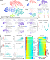
Single-cell RNA-seq analysis identifies 5 patterns of differential gene expression upon Nkx2-1 deletion. (A) Seurat t-distributed stochastic neighbor embedding (tSNE) plots of sorted epithelial cells from littermate lungs (merged and split views in the 2 Left columns). Most epithelial cells in the mutant lung do not express Nkx2-1 (Top in the Rightmost column). Color-coded cell types are tabulated and identified using the following markers: control AT1, Aqp5; mutant AT1, Nkx2-1 negative (early mutant AT1 is closer to control AT1 spatially in the tSNE plot); proliferative, Mki67 (B); AT2/AT1, coexpression of Lamp3 and Aqp5; AT2, Lamp3; club, Scgb1a1; and ciliated, Foxj1. See SI Appendix, Fig. S5D. (B) Representative genes for each pattern of differential gene expression. Early up-/down-regulated genes exhibit changes in the early mutant AT1 cluster, whereas late up-/down-regulated genes exhibit changes in the late mutant AT1 cluster. The biphasic genes are first up-regulated and then down-regulated as control AT1 cells transition to early and then late mutant AT1 cells. (C) Monocle pseudotemporal analysis of AT1 cells from control and mutant lungs, excluding Mki67 cells. (D) Pseudotemporal changes of representative genes for the 5 patterns of differential gene expression. Vertical dashes demarcate loss of Nkx2-1 expression (also in E). (E) Heatmap of pseudotime-dependent gene clusters from the top 1,000 differentially expressed genes. Nkx2-1 loss is marked by vertical dashes. A total of 24 out of 27 AT1 cell-specific genes (blue gene names) are in the down-regulated groups.
Fig. 7.
Fig. 7.
NKX2-1 binds near AT1 and down-regulated genes. (A) NKX2-1 ChIP peaks (gray arrows) near genes representative of the 5 patterns of differential gene expression (Fig. 6) using wild-type P10 lungs and 2 independent antibodies (Ab1, Abcam ab133737; Ab2, Millipore 07-601). (B) Average (per gene) NKX2-1 ChIP peak profiles that are color coded as in A and tabulated in the order of peak height. The 27-AT1 gene group has the highest average binding. The whole genome (all; 23,793 genes) serves as a reference for background binding. Asterisks: P < 0.0001; n.s., not significant (χ2 test). (C) A model summarizing the 4 deletion models and showing that Nkx2-1 is required for the specification, development, and maintenance of AT1 cells (green).

Similar articles

Cited by

References

    1. Morrisey E. E., Hogan B. L., Preparing for the first breath: Genetic and cellular mechanisms in lung development. Dev. Cell 18, 8–23 (2010). - PMC - PubMed
    1. Weibel E. R., On the tricks alveolar epithelial cells play to make a good lung. Am. J. Respir. Crit. Care Med. 191, 504–513 (2015). - PubMed
    1. Weibel E. R., The mystery of “non-nucleated plates” in the alveolar epithelium of the lung explained. Acta Anat. (Basel) 78, 425–443 (1971). - PubMed
    1. Yang J., Chen J., Developmental programs of lung epithelial progenitors: A balanced progenitor model. Wiley Interdiscip. Rev. Dev. Biol. 3, 331–347 (2014). - PMC - PubMed
    1. Yang J., et al. , The development and plasticity of alveolar type 1 cells. Development 143, 54–65 (2016). - PMC - PubMed

Publication types

Substances