. 2019 Oct 8;116(41):20689-20699.
doi: 10.1073/pnas.1902958116. Epub 2019 Sep 23.

Parkinson's disease-associated iPLA2-VIA/ PLA2G6 regulates neuronal functions and α-synuclein stability through membrane remodeling

Affiliations

Parkinson's disease-associated iPLA2-VIA/ PLA2G6 regulates neuronal functions and α-synuclein stability through membrane remodeling

Akio Mori et al. Proc Natl Acad Sci U S A. .

Abstract

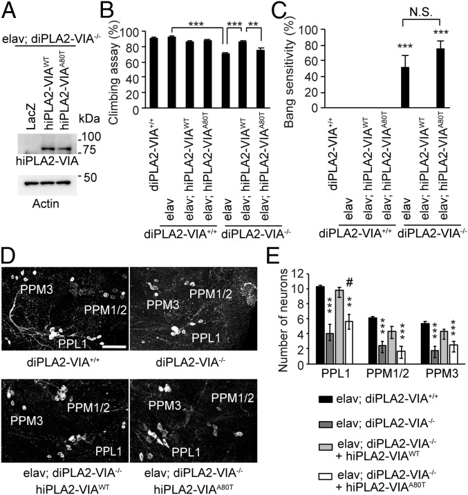
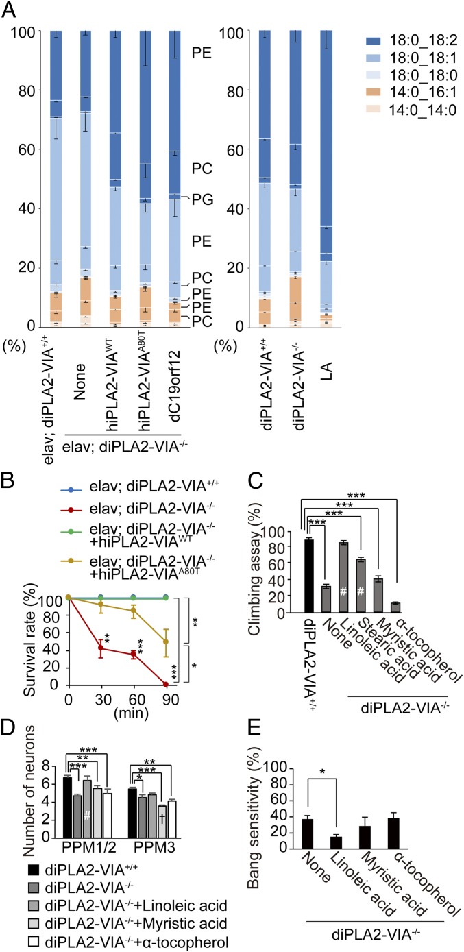
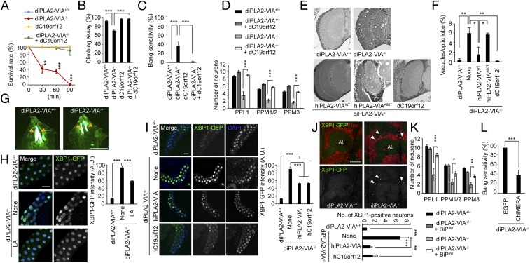
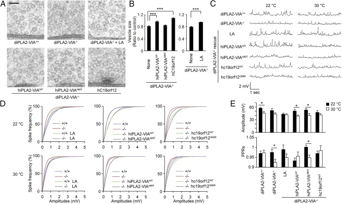
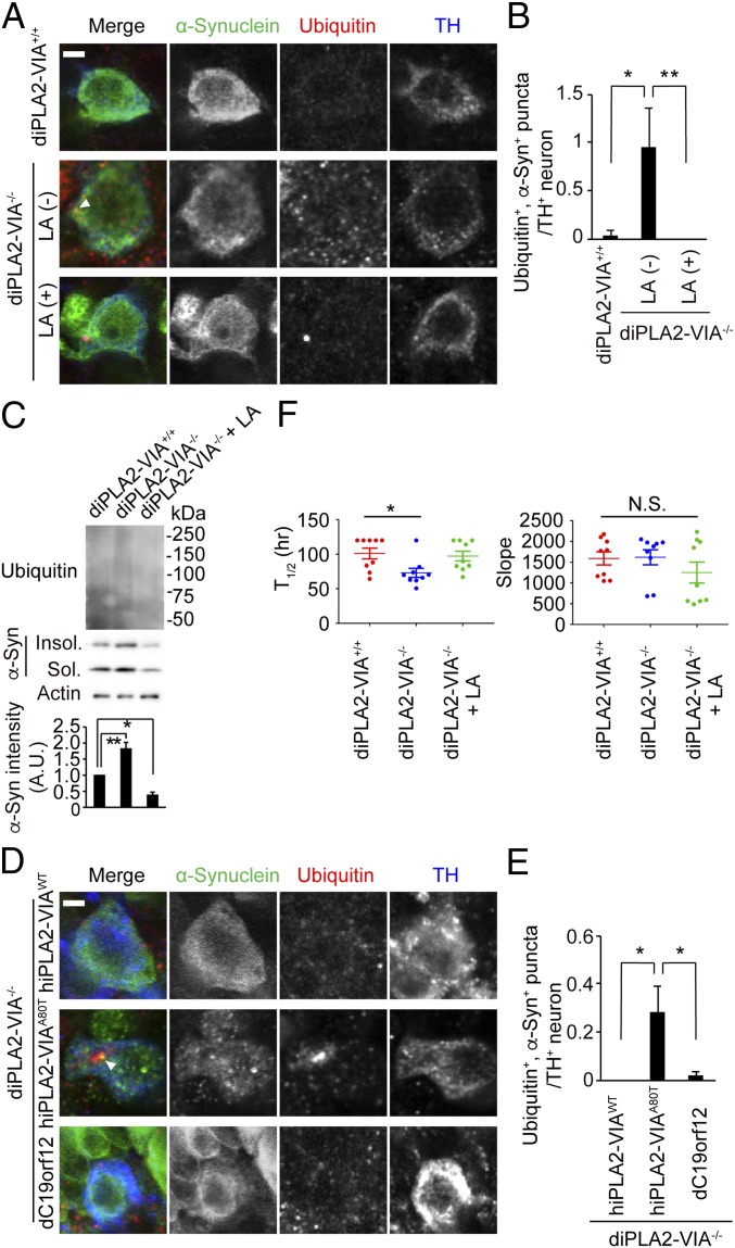
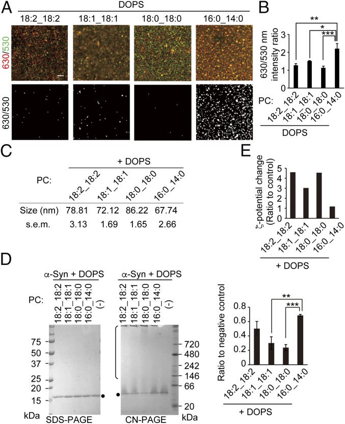
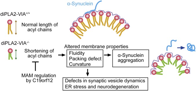
Mutations in the iPLA2-VIA/PLA2G6 gene are responsible for PARK14-linked Parkinson's disease (PD) with α-synucleinopathy. However, it is unclear how iPLA2-VIA mutations lead to α-synuclein (α-Syn) aggregation and dopaminergic (DA) neurodegeneration. Here, we report that iPLA2-VIA-deficient Drosophila exhibits defects in neurotransmission during early developmental stages and progressive cell loss throughout the brain, including degeneration of the DA neurons. Lipid analysis of brain tissues reveals that the acyl-chain length of phospholipids is shortened by iPLA2-VIA loss, which causes endoplasmic reticulum (ER) stress through membrane lipid disequilibrium. The introduction of wild-type human iPLA2-VIA or the mitochondria-ER contact site-resident protein C19orf12 in iPLA2-VIA-deficient flies rescues the phenotypes associated with altered lipid composition, ER stress, and DA neurodegeneration, whereas the introduction of a disease-associated missense mutant, iPLA2-VIA A80T, fails to suppress these phenotypes. The acceleration of α-Syn aggregation by iPLA2-VIA loss is suppressed by the administration of linoleic acid, correcting the brain lipid composition. Our findings suggest that membrane remodeling by iPLA2-VIA is required for the survival of DA neurons and α-Syn stability.

Keywords: Drosophila; ER stress; Parkinson’s disease; lipids; α-synuclein.


Conflict of interest statement

The authors declare no conflict of interest.

Figures

Fig. 1.
Fig. 1.
Loss of iPLA2-VIA leads to DA neurodegeneration. (A) Complementary expression of hiPLA2-VIA in diPLA2-VIA/ flies. Expression of hiPLA2-VIA was determined by Western blot with anti-hiPLA2-VIA. LacZ and Actin served as a mock transgene and a loading control, respectively. (B and C) Defects in motor activity and the bang-sensitive seizure phenotype of diPLA2-VIA/ flies are fully rescued by hiPLA2-VIA WT but not A80T. The climbing assay (n = 20 trials in 20 male flies) and the bang test (n = 5 trials in 10 male flies) were performed using 5-d-old flies, and data are represented as the mean ± SEM (**P < 0.01 and ***P < 0.001 vs. any other groups in C) by 1-way ANOVA with the Tukey–Kramer test. N.S., not significant. (D and E) DA neurons decrease in diPLA2-VIA/ flies, which is alleviated by hiPLA2-VIA expression. (D) The PPL1, PPM1/2, and PPM3 cluster DA neurons were visualized in 20-d-old adult brain tissues by an anti-TH antibody. (Scale bar, 50 μm.) (E) The number of TH-positive neurons is graphed. Data are represented as the mean ± SEM (n = 14 to 21 flies; **P < 0.01 and ***P < 0.001 vs. diPLA2-VIA+/+; #P < 0.05 vs. hiPLA2-VIAWT by 1-way ANOVA with the Tukey–Kramer test). Transgenes were driven by the pan-neuronal elav-GAL4 driver.
Fig. 2.
Fig. 2.
hiPLA2-VIA introduction and linoleic acid supplementation reverse the altered lipid composition and neuronal phenotypes. (A) Higher rate of shorter acyl chains in diPLA2-VIA/ and diPLA2-VIA/ flies expressing hiPLA2-VIA A80T. The phospholipid composition of the diPLA2-VIA/ brain is corrected by dC19orf12 expression or an LA-rich diet. The compositions of shorter acyl chains (14:0_14:0 and 14:0_16:1) and longer acyl chains (18:0_18:0, 18:0_18:1, and 18:0_18:2) in phospholipids and the composition of the phospholipid head groups from 20-d-old adult fly brain tissues are shown as a bar graph (mean of 3 to 4 independent samples). (B) diPLA2-VIA/ flies exhibit sensitivity to high temperature. Twenty-day-old adult flies were incubated at 37 °C, and the percentage of survivors was calculated at the indicated time points (mean ± SEM, 3 trials with 10 flies; *P = 0.011, **P < 0.01, and ***P < 0.001 at each time point by 1-way ANOVA with the Tukey–Kramer test). (C) An LA-rich diet improves motor ability in diPLA2-VIA/ flies. The climbing assay was performed by 3-d-old adult flies raised on normal instant fly food or an instant fly food containing FAs with different lengths of acyl chains or α-tocopherol (mean ± SEM, n = 10 to 15 flies, 20 trials; ***P < 0.001 by Dunnett’s test; #P < 0.001 vs. diPLA2-VIA/ [none] by 1-way ANOVA with the Tukey–Kramer test). (D) An LA-rich diet rescues DA neuron loss caused by lack of diPLA2-VIA. The number of DA neurons in each cluster in 20-d-old adult flies (mean ± SEM, n = 11 to 23; *P < 0.05, **P < 0.01, and ***P < 0.001 by Dunnett’s test; P < 0.005 and #P < 0.001 vs. diPLA2-VIA/ by 1-way ANOVA with the Tukey–Kramer test). (E) The bang-sensitive seizure-paralysis phenotype of 5-d-old diPLA2-VIA/ flies is rescued by LA supplementation. Data are represented as the mean ± SEM (n = 10 male flies, 4 trials; *P < 0.05 by Dunnett’s test).
Fig. 3.
Fig. 3.
Neurodegeneration of diPLA2-VIA/ flies is rescued by MPAN-associated C19orf12. (A) Neuronal expression of the MPAN-associated gene dC19orf12 improves sensitivity to high temperature. The assay was performed as described in Fig. 2B (mean ± SEM, 3 trials with 10 flies at 20 d old; **P < 0.001 and ***P < 0.0001 by 1-way ANOVA with the Tukey–Kramer test). (B and C) Neuronal expression of dC19orf12 rescues motor disability and bang-sensitive seizures in diPLA2-VIA/ flies. The climbing (mean ± SEM, n = 10 to 15 flies, 20 trials) and bang sensitivity (mean ± SEM, n = 30 to 40 flies) assays were performed with 3-d-old adult flies (***P < 0.0001 by 1-way ANOVA with the Tukey–Kramer test). (D) dC19orf12 rescues DA neuron loss caused by lack of diPLA2-VIA. The number of DA neurons in the indicated clusters is graphed (mean ± SEM, n = 12 to 22 flies at 20 d old; ***P < 0.0001 by 1-way ANOVA with the Tukey–Kramer test). (E and F) Expression of hiPLA2-VIA WT and dC19orf12 suppresses the vacuolation of the diPLA2-VIA/ fly brain. (E) Light-microscopic examination of brain tissue sections from 20-d-old flies with the indicated genotypes. (F) The percentage of total vacuole areas accounting for the optic lobe regions is graphed (mean ± SEM, n = 3 flies; *P < 0.05 and **P < 0.01 by 1-way ANOVA with the Tukey–Kramer test). (G) Poor development of salivary glands (arrows) caused by lack of diPLA2-VIA. The salivary glands at the third-instar larval stage were visualized by ER-mAG1. (Scale bar, 1 mm.) (H and I) ER stress in the salivary glands of the diPLA2-VIA/ larvae is improved by LA (1 μL/mL) treatment or by the expression of hiPLA2-VIA or dC19orf12. XBP1-GFP and DAPI images in the salivary glands of third-instar larvae with the indicated genotypes. (Scale bars, 50 µm.) Graphs represent XBP1-GFP signal intensities with the indicated genotypes (n = 142 to 526 cells in 3 to 12 larvae; ***P < 0.001 by 1-way ANOVA with the Tukey–Kramer test). (J) Neuronal ER stress caused by iPLA2-VIA loss is relieved by the expression of hiPLA2-VIA or dC19orf12. Images show XBP1-GFP and Elav from an anterior view in the brains of 20-d-old flies. (Scale bar, 50 µm.) Arrowheads indicate XBP1- and Elav-positive signals. Elav was used as a neuron-specific nuclear marker. AL, antennal lobe. Graph represents the number of XBP1-GFP–positive neurons per brain (n = 9 to 13 flies; **P < 0.005 and ***P < 0.001 by 1-way ANOVA with the Tukey–Kramer test). (K) DA neuron loss caused by lack of diPLA2-VIA is improved by WT BiP (BiPWT) in diPLA2-VIA/ flies at 20 d old. The number of DA neurons in the indicated clusters is graphed (mean ± SEM, n = 7 to 15 flies; *P < 0.01, **P < 0.001, and ***P < 0.0001 by 1-way ANOVA with the Tukey–Kramer test). (L) The bang-sensitive seizure phenotype is rescued by ChiMERA but not EGFP in diPLA2-VIA/ flies at 20 d old. Data are represented as the mean ± SEM (n = 17 EGFP and n = 33 ChiMERA flies; ***P < 0.001 by 2-tailed Student’s t test).
Fig. 4.
Fig. 4.
Altered acyl-chain composition affects synaptic vesicle size. (A) Ultrastructure of the synaptic vesicles at the active zones in third-instar larval motor neurons with the indicated genotypes. (Scale bar, 100 nm.) (B) The ratio of synaptic vesicle diameter to diPLA2-VI+/+ is graphed (mean ± SEM, n = 130 to 150 from 3 third-instar larvae; ***P < 0.0001 by 1-way ANOVA with the Tukey–Kramer test [Left] and by 2-tailed Student’s t test [Right]). (C) Representative spontaneous mEJP traces in the third-instar larval NMJ at 22 and 30 °C. (D) Cumulative distribution of mEJP amplitude at 22 and 30 °C over 60 s (n = 7 to 11 third-instar larvae per genotype). (E) EJP amplitude evoked by 1.5 to 2 V electrical stimulation at 22 and 30 °C and paired-pulse ratio (PPR) as a ratio of EJP amplitudes to paired electrical stimulations (50-ms interval) at 22 and 30 °C (n = 7 to 9 third-instar larvae per genotype; *P < 0.05 by 2-tailed Student’s t test).
Fig. 5.
Fig. 5.
Alteration of phospholipid composition by diPLA2-VIA loss promotes α-Syn aggregation. (AC) α-Syn accumulation by diPLA2-VIA loss is relieved by the LA diet. (A) Brain tissues from 15-d-old flies were stained with the indicated antibodies. An arrowhead indicates ubiquitin- and α-Syn–positive punctum. (Scale bar, 2 μm.) (B) Ubiquitin- and α-Syn–positive puncta in TH-positive neurons are graphed (n = 10 to 15 neurons from 6 flies at 15 d old per sample; *P < 0.05 and **P < 0.01 by 1-way ANOVA with the Tukey–Kramer test). (C) Sarkosyl-soluble and -insoluble fractions from brain tissues from 20-d-old flies were analyzed by Western blot. The band intensities of α-Syn normalized to those of actin are graphed (n = 4; *P < 0.05 and **P < 0.01 by Dunnett’s test). (D and E) α-Syn accumulation caused by diPLA2-VIA loss is suppressed by hiPLA2-VIA or dC19orf12. (D) Brain tissues from 15-d-old flies were stained with the indicated antibodies. An arrowhead indicates ubiquitin- and α-Syn–positive punctum. (Scale bar, 2 μm.) (E) Ubiquitin- and α-Syn–positive puncta in TH-positive neurons are graphed (n = 10 to 15 neurons from 6 flies at 15 d old per sample; *P < 0.05 by 1-way ANOVA with the Tukey–Kramer test). (F) diPLA2-VIA loss promotes the seeding activity of α-Syn. Brain lysates prepared from 15-d-old flies neuronally expressing α-Syn were subjected to RT-QUIC. Graphs indicate the time when TdT fluorescence reached an intensity of 165,000 RFU (Left, t1/2) and the slope obtained by differential processing of plot curves (Right), respectively. When TdT fluorescence did not reach 165,000 RFU before 120 h, the value of t1/2 was defined as 120 (n = 3 replicates from 3 flies per sample; *P < 0.05 by 1-way ANOVA with the Tukey–Kramer test). N.S., not significant; RFU, relative fluorescence units.
Fig. 6.
Fig. 6.
A shorter acyl-chain composition weakens α-Syn association with liposomes. (A and B) Lipid packing status of SUVs. (A) SUVs containing 30% DOPS and 70% of the indicated phospholipids were incubated with 250 nM di-4-ANEPPDHQ, and the fluorescence intensities at 630 and 530 nm were imaged (Upper). The 630-nm signal divided by the thresholded 530-nm signal intensity is also shown (Lower). (Scale bar, 2 μm.) (B) The 630/530-nm intensity ratios are graphed (n = 3 to 4; *P < 0.05, **P < 0.01, and ***P < 0.005 by 1-way ANOVA with the Tukey–Kramer test). (C) Diameter (in nanometers) of SUVs containing 30% DOPS and 70% of the indicated phospholipids (mean ± SEM; 3 trials). (D) α-Syn does not bind to SUVs containing shorter acyl chains. Five micromolar recombinant α-Syn incubated with 500 µM SUV as prepared in AC for 10 min was separated by clear-native PAGE (CN-PAGE) or SDS/PAGE. Dots and open bracket indicate a monomer form of α-Syn and slower-migrating forms of α-Syn due to the interaction with SUV, respectively. Graph represents the ratios of α-Syn monomer signals to a control without SUV on CN-PAGE, normalized to α-Syn signals that appeared on SDS/PAGE as inputs (n = 3; **P < 0.01 and ***P < 0.001 by Dunnett’s test). (E) ζ Potentials of SUVs with recombinant α-Syn are indicated as a ratio to that of control liposomes without proteins.
Fig. 7.
Fig. 7.
Working hypothesis. Loss of iPLA2-VIA causes the shortening of phospholipid acyl chains in the brain, which alters membrane fluidity, lipid packing, and curvature. α-Syn affinity to the synaptic membrane is weakened by altered phospholipid properties, leading to α-Syn aggregation. These altered membrane properties also induce ER stress, affect the dynamics of SVs, and provoke abnormal neurotransmission, which manifests as the bang-sensitivity phenotype. Correction of the membrane composition by dietary intake of FAs or enhanced MAM integrity by C19orf12 rescues neurodegeneration and α-Syn aggregation.

Similar articles

Cited by

References

    1. Kalia L. V., Lang A. E., Parkinson’s disease. Lancet 386, 896–912 (2015). - PubMed
    1. Auluck P. K., Caraveo G., Lindquist S., α-Synuclein: Membrane interactions and toxicity in Parkinson’s disease. Annu. Rev. Cell Dev. Biol. 26, 211–233 (2010). - PubMed
    1. Jao C. C., Hegde B. G., Chen J., Haworth I. S., Langen R., Structure of membrane-bound alpha-synuclein from site-directed spin labeling and computational refinement. Proc. Natl. Acad. Sci. U.S.A. 105, 19666–19671 (2008). - PMC - PubMed
    1. Bozek K., et al. , Organization and evolution of brain lipidome revealed by large-scale analysis of human, chimpanzee, macaque, and mouse tissues. Neuron 85, 695–702 (2015). - PubMed
    1. Antonny B., Vanni S., Shindou H., Ferreira T., From zero to six double bonds: Phospholipid unsaturation and organelle function. Trends Cell Biol. 25, 427–436 (2015). - PubMed

Publication types

MeSH terms