. 2019 Oct 8;116(41):20612-20622.
doi: 10.1073/pnas.1805404116. Epub 2019 Sep 23.

RNA ligation precedes the retrotransposition of U6/LINE-1 chimeric RNA

Affiliations

RNA ligation precedes the retrotransposition of U6/LINE-1 chimeric RNA

John B Moldovan et al. Proc Natl Acad Sci U S A. .

Abstract

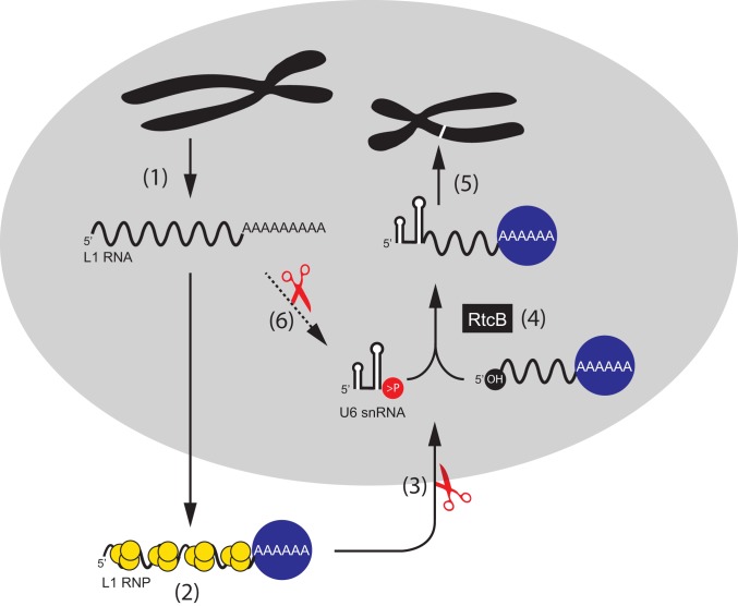
Long interspersed element-1 (LINE-1 or L1) amplifies via retrotransposition. Active L1s encode 2 proteins (ORF1p and ORF2p) that bind their encoding transcript to promote retrotransposition in cis The L1-encoded proteins also promote the retrotransposition of small-interspersed element RNAs, noncoding RNAs, and messenger RNAs in trans Some L1-mediated retrotransposition events consist of a copy of U6 RNA conjoined to a variably 5'-truncated L1, but how U6/L1 chimeras are formed requires elucidation. Here, we report the following: The RNA ligase RtcB can join U6 RNAs ending in a 2',3'-cyclic phosphate to L1 RNAs containing a 5'-OH in vitro; depletion of endogenous RtcB in HeLa cell extracts reduces U6/L1 RNA ligation efficiency; retrotransposition of U6/L1 RNAs leads to U6/L1 pseudogene formation; and a unique cohort of U6/L1 chimeric RNAs are present in multiple human cell lines. Thus, these data suggest that U6 small nuclear RNA (snRNA) and RtcB participate in the formation of chimeric RNAs and that retrotransposition of chimeric RNA contributes to interindividual genetic variation.

Keywords: LINE-1; RNA ligation; RtcB; U6 snRNA; retrotransposon.


Conflict of interest statement

Conflict of interest statement: J.V.M. is an inventor on patent US6150160, is a paid consultant for Gilead Sciences and a privately held company founded by Flagship Pioneering, and is on the American Society of Human Genetics Board of Directors.

Figures

Fig. 1.
Fig. 1.
Chimeric U6/L1 RNA is generated in HeLa cells transfected with human L1 expression constructs. (A) Schematics of wild-type and mutant L1s. Gray rectangles represent 5′ UTR, inter-ORF space, and 3′ UTR, respectively; yellow rectangle, ORF1; blue rectangle, ORF2. L1s were cloned into the pCEP4 mammalian expression vector. A cytomegalovirus immediate early promoter (CMV, black rectangle) that augments L1 expression and an SV40 polyadenylation signal (light blue rectangle, pA) flank the L1. The pJM101/L1.3Δneo plasmid expresses an active human L1 (L1.3). The pJM108/L1.3Δneo, pJM105/L1.3Δneo, and pJBM119/L1.3Δneo plasmids express versions of L1.3 that contain mutations that render them unable to retrotranspose; the approximate locations of the respective mutations are indicated in the schematic. (B) Rationale of the RT-PCR experiments used to detect U6/L1 chimeric RNAs. HeLa cells were transfected with L1 expression plasmids, total cellular RNA was extracted ∼48 h posttransfection, and cDNAs were synthesized using an oligo-dT primer. Nested PCR was carried out using primers complementary to sequences within U6 and the 3′ end of the L1 construct (U6s1 and SV40as, then U6s2 and 3UTRas3). (C) Results from a representative RT-PCR experiment. The transfected L1 construct is indicated above each lane of the agarose gel image. Each lane contains a single biological replicate. Lane 1, HeLa UTF (untransfected HeLa cells); lanes 2 and 3, HeLa transfected with pJM101/L1.3Δneo; lanes 4 and 5, HeLa transfected with pJM105/L1.3Δneo; lanes 6 and 7, HeLa transfected with pJBM119/L1.3Δneo; lanes 8 and 9, HeLa transfected with pJM108/L1.3Δneo; lanes 10 and 11, H2O PCR controls. Molecular weight standards (in bp) are shown in the first and last gel lanes. At least 3 independent biological replicates were conducted for each transfection condition. (D) Structures of 38 U6/L1 chimeric RNAs found in transfected HeLa cells. U6/L1 RNA chimera sequences contain the 3′ terminus of U6 snRNA cDNA (white arrow) ending in ∼4 to 6 thymidine nucleotides (Tn) conjoined to a variable 5′−truncated L1. A schematic of the full-length L1.3 sequence is represented at the top of the schematic. The horizontal black lines indicate the approximate length of L1 sequence conjoined to the U6 poly(T) tract. The 5′-most U6/L1 junction occurred at L1.3 nucleotide position 4387.
Fig. 2.
Fig. 2.
Purified recombinant RtcB ligates U6 RNA to L1 RNA in vitro. (A) The rationale of the U6/L1 in vitro ligation experiment. A synthetic human U6 RNA containing a 2′,3′-cyclic phosphate (>P, red circle) and a synthetic L1 RNA (blue font) containing a 5′-OH (black circle) were generated using a ribozyme-based in vitro transcription reaction. U6 and L1 RNAs were splinted with a cDNA oligonucleotide (DNA splint, italics), and then the resultant RNA/DNA hybrid was incubated with purified RtcB (black rectangle, white font) for 1 h. The reaction was then treated with DNase I, the RNA was purified, and cDNAs were synthesized using the SV40as oligonucleotide primer. RT-PCR reactions using nested primers (U6s1 and SV40as, then U6s2 and 3UTRas3) were used to detect U6/L1 chimeric cDNAs. (B) Schematic representations of the synthetic RNAs used in in vitro experiments. The in vitro transcribed U6 RNA ends in 4 uridine ribonucleotides and contains a 2′,3′-cyclic phosphate (U6 > P) or a 3′-OH (U6-OH). The in vitro transcribed L1 RNA consists of pJM101/L1.3Δneo sequence (nt positions 5752 to 6087) and contains a 5′-OH (OH-L1) or a 5′-triphosphate (P-L1). (C) Results from the in vitro U6/L1 ligation reactions. The constituents of U6/L1 ligation reactions are indicated above each gel lane (+) of the agarose gel image. An asterisk (*) indicates that RtcB was heat treated at 95 °C for 10 min prior to adding it to the reaction. No RT, no RT control; H2O, water PCR controls. DNA size markers (in bp) are shown to the left of the gel image. The predicted position of the 305-bp U6/L1 RT-PCR product is noted on the left side of the gel image (white arrow, red font). (D) Summary of results from product characterization experiments. Column 1, synthetic RNAs used in the reaction; column 2, number of RT-PCR products characterized for each reaction condition; column 3, number of RT-PCR products that correspond to the full-length ligation product; column 4, number of RT-PCR products that contain a variably 5′-truncated L1 sequence; column 5, number of putative RT-PCR artifact products. Each in vitro experiment was repeated 3 independent times and yielded similar results.
Fig. 3.
Fig. 3.
HeLa cell nuclear extracts mediate the ligation of U6 and L1 RNAs. (A) Results from U6/L1 ligation reactions using HeLa cell nuclear extracts. The ligated U6/L1 RNA was purified from ligation reactions and analyzed using RT-PCR and agarose gel electrophoresis. The constituents of U6/L1 ligation reactions are indicated above each lane (+) of the representative agarose gel image. An asterisk (*) indicates that the HeLa cell nuclear extract was heat treated at 95 °C for 10 min prior to adding it to the reaction. No RT, no RT control; H2O, water PCR controls. DNA size markers (in bp) are shown to the left of the gel image. The predicted position of the 305-bp U6/L1 RT-PCR product is noted on the left side of the gel image (white arrow, red font). (B) Summary of results of ligation reactions using HeLa cell extracts. Column 1, RNAs used in the reaction; column 2, number of RT-PCR products characterized for each reaction condition; column 3, number of RT-PCR products that correspond to the full-length ligation product; column 4, number of RT-PCR products that contain a variably 5′-truncated L1 sequence; column 5, number of putative RT-PCR artifact products. (C) Structures of U6/L1 chimeric RNAs containing 5′-variably truncated L1 sequences. A schematic of the L1 fragment used as a template for the in vitro transcription reaction is represented at the top. The horizontal black lines indicate the approximate length of the L1 sequence conjoined to the U6 poly(T) tract. U6/L1 chimeric RNAs were isolated from 19 independent experiments. (D) Depletion of RtcB protein expression in HeLa cell nuclear extracts. Western blot images depicting RtcB expression (green arrow) in HeLa nuclear extracts. Extract sources are indicated above each lane (HeLa indicates untransfected HeLa extracts). Each lane represents an independent biological replicate. The Western blot experiment was done twice. Nucleolin (NCL) (red arrow) was used as a loading control. Approximate molecular weights are indicated to the left of the gel image. (E) Depletion of RtcB affects U6/L1 ligation efficiency in HeLa extracts. The x axis indicates the experimental condition. The y axis indicates the normalized U6/L1 ligation efficiency. Ligation efficiencies were normalized to untransfected HeLa-JVM extracts, which are set to 1. The ligation efficiency value represents the average of 6 independent RT-qPCR experiments. Error bars indicate SDs. Two-tailed t tests were used to determine significance. An asterisk (*) indicates P value < 0.05; n.s., not significant.
Fig. 4.
Fig. 4.
RNA-seq detection of endogenous U6/L1 chimeric RNAs in human cell lines. (A) Rationale of the RNA-seq experiments. Step 1: Ribosome-depleted RNA was fragmented to ∼190 nucleotides and subjected to 100-bp paired-end DNA sequencing. Step 2: RNA-seq read pairs (arrows) were aligned to a repeat masked version human reference genome (HGR/build Grch38), which contained “spiked-in” copies of a single U6 (white rectangle) and single L1.3 (blue rectangle) sequence. RNA-seq reads that did not map to U6 or L1 were discarded from subsequent analyses. Step 3: Overlapping U6/L1 read pairs were merged to determine the U6/L1 junction sequences. U6/L1 read pairs that contained a gap (i.e., “no overlap”) were discarded from subsequent analyses. Step 4: Overlapping U6/L1 junctions were aligned to the unmasked HGR. If the U6/L1 junction mapped to the HGR with >90% accuracy, it was designated as an “aligned” read. U6/L1 junctions that failed to map to the HGR with at least 90% accuracy were designated as “non-aligned” reads. (B) Structures of RNA-seq U6/L1 junctions. A schematic of a full-length RC-L1 is indicated at the top. The general structure of a U6/L1 chimeric junction sequence consists of the 3′ end of a U6 snRNA cDNA sequence ending in ∼4 to 8 thymidine nucleotides (left side of figure; white arrow ending in Tn) conjoined to a variably 5′−truncated L1 sequence. Two independent RNA-seq libraries were generated from HeLa-JVM, H9, NPC, and PA-1 cells, respectively (squares and triangles, respectively). One RNA-seq library was generated from HeLa-HA cells. Red horizontal lines, HeLa-JVM; green horizontal lines, HeLa-HA; yellow horizontal lines, PA-1; black horizontal lines, H9; blue horizontal lines, human NPCs. Each horizontal dashed line represents a single U6/L1 junction RNA-seq merged sequence read. The triangle or square at the left end of the horizontal dashed lines indicates the approximate location of the U6/L1 junction point relative to L1.3. The top set of dashed lines represent 16 U6/L1 junction sequences that mapped (“aligned”) to the HGR. The bottom set of dashed lines represent 33 U6/L1 junction sequences that did not map (“non-aligned”) to the HGR. These 33 U6/L1 junctions contained a copy of U6 conjoined to an L1 present in the same transcriptional orientation. The remaining 4 U6/L1 chimeras that did not map to the HGR (SI Appendix, Table S6) contained a copy of U6 conjoined to an L1 present in the opposite transcriptional orientation.
Fig. 5.
Fig. 5.
A model of U6/L1 RNA pseudogene formation. Following transcription, L1 RNA (black wavy line) is exported to the cytoplasm (step 1). L1 RNA is translated, and the L1 proteins, ORF1p (yellow circles) and ORF2p (blue circle) bind to their encoding L1 RNA to form an L1 ribonucleoprotein particle (RNP) (step 2). After RNP formation, the L1 RNA is cleaved by an unidentified endoribonuclease to generate 5′-truncated L1 RNA with a 5′-OH (step 3). RtcB, or a related ligase, ligates U6 snRNA to 5′-truncated L1 RNA (step 4). The resultant chimeric U6/L1 RNA is inserted into genomic DNA by TPRT (in cis), which results in the formation of a U6/L1 chimeric pseudogene (white band on black chromosome) (step 5). It is possible that L1 RNA could also be processed by nuclease activity (red scissors) and ligated to U6 while still in the nucleus (step 6). In this scenario, it is possible the chimeric U6/L1 RNA could undergo retrotransposition by trans-complementation.

References

    1. Lander E. S., et al. ; International Human Genome Sequencing Consortium , Initial sequencing and analysis of the human genome. Nature 409, 860–921 (2001). Erratum in: Nature411, 720 (2001) and Nature412, 565 (2001). - PubMed
    1. Smit A. F., The origin of interspersed repeats in the human genome. Curr. Opin. Genet. Dev. 6, 743–748 (1996). - PubMed
    1. Grimaldi G., Skowronski J., Singer M. F., Defining the beginning and end of KpnI family segments. EMBO J. 3, 1753–1759 (1984). - PMC - PubMed
    1. Scott A. F., et al. , Origin of the human L1 elements: Proposed progenitor genes deduced from a consensus DNA sequence. Genomics 1, 113–125 (1987). - PMC - PubMed
    1. Ostertag E. M., Kazazian H. H. Jr, Twin priming: A proposed mechanism for the creation of inversions in L1 retrotransposition. Genome Res. 11, 2059–2065 (2001). - PMC - PubMed

Publication types

LinkOut - more resources