. 2019 Oct 8;116(41):20482-20488.
doi: 10.1073/pnas.1905722116. Epub 2019 Sep 23.

Cell division rates decrease with age, providing a potential explanation for the age-dependent deceleration in cancer incidence

Affiliations

Cell division rates decrease with age, providing a potential explanation for the age-dependent deceleration in cancer incidence

Cristian Tomasetti et al. Proc Natl Acad Sci U S A. .

Abstract

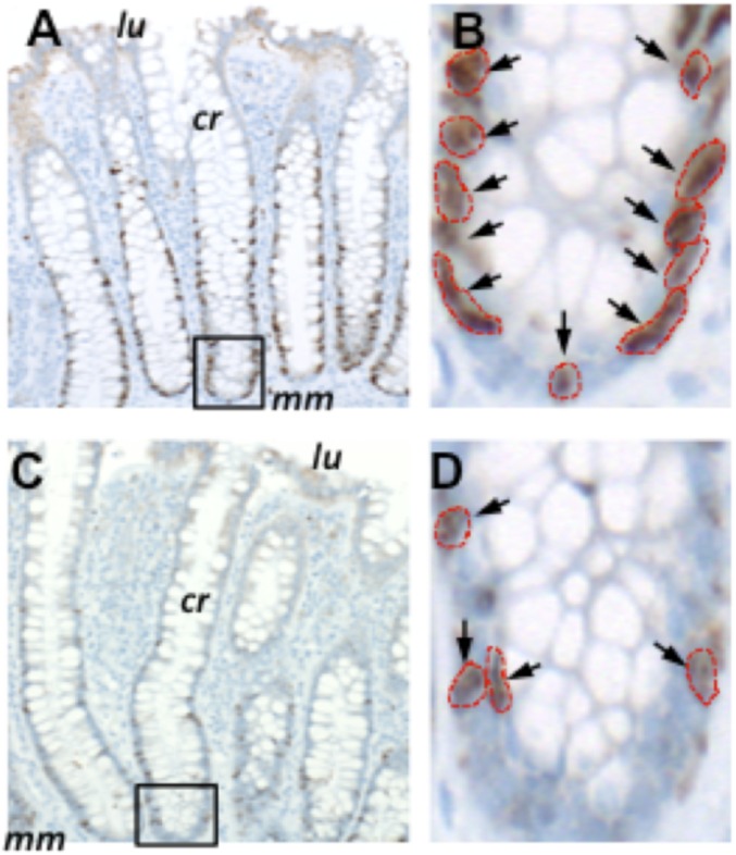
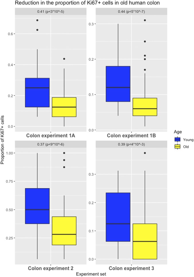
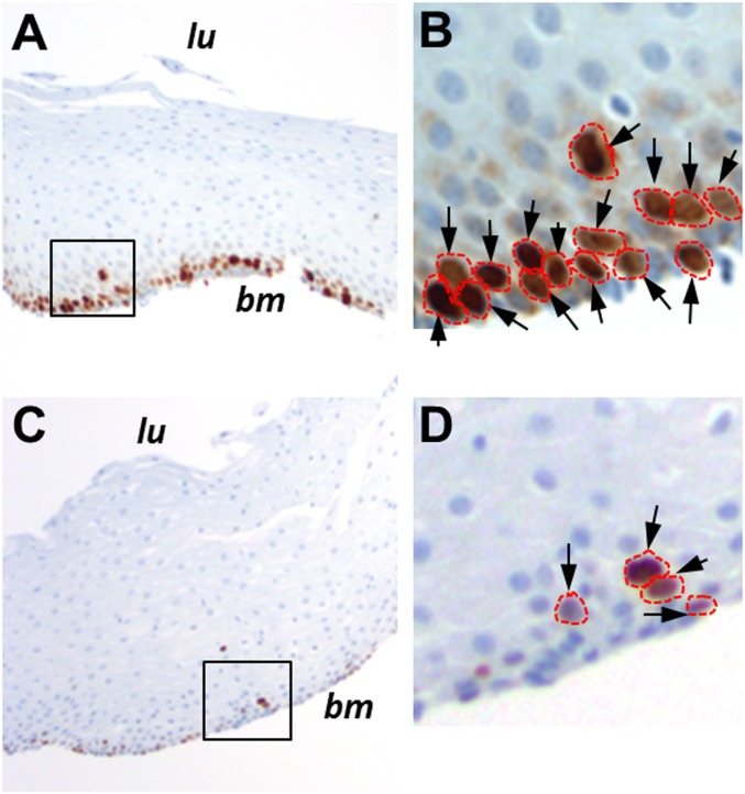
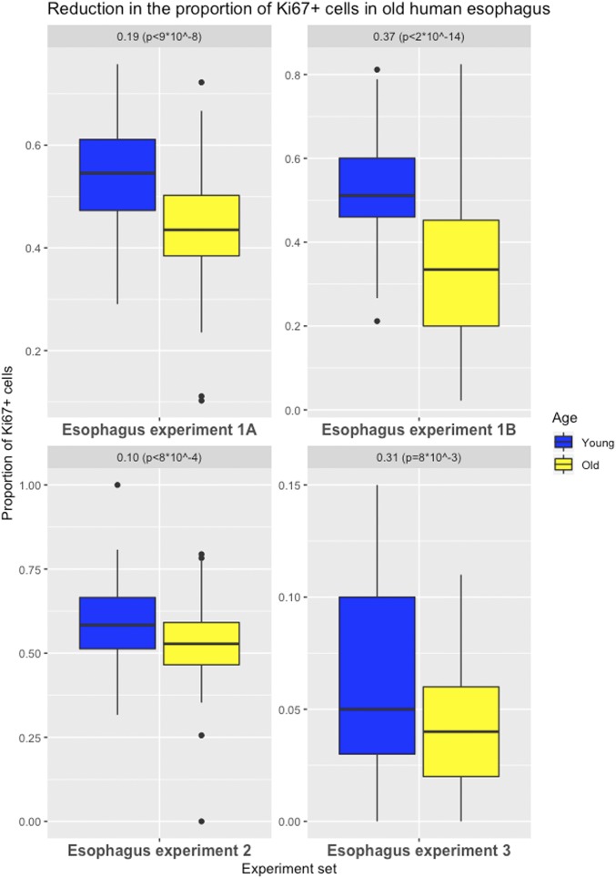
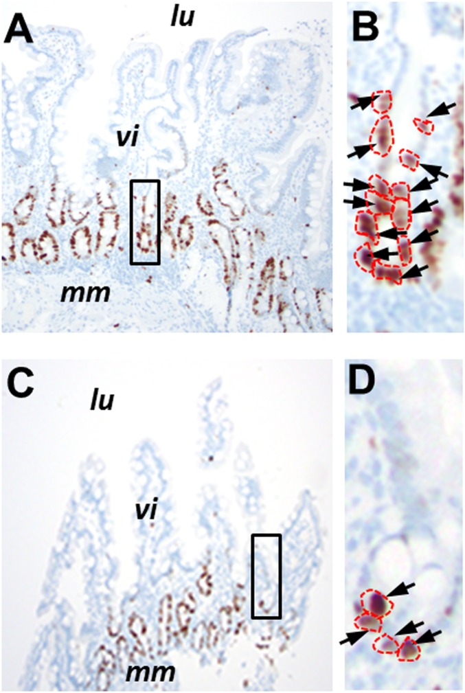
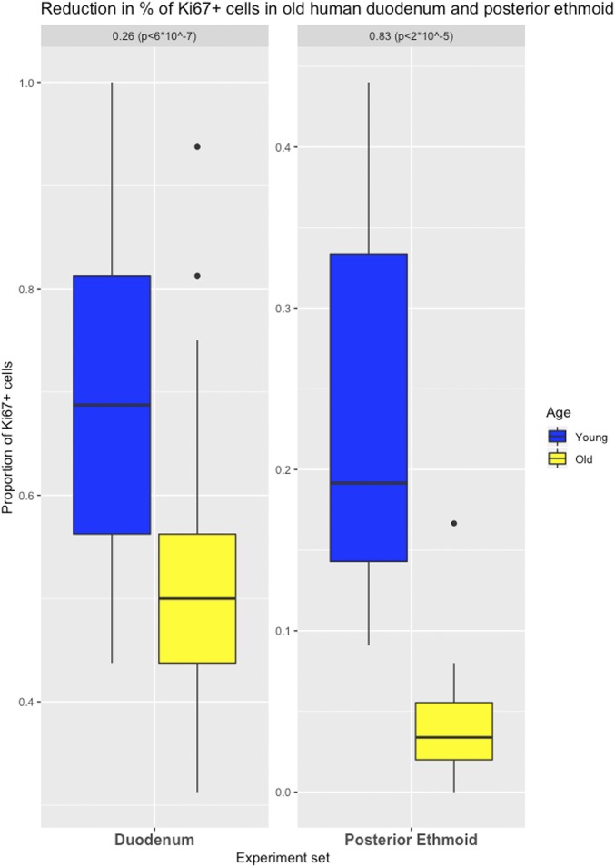
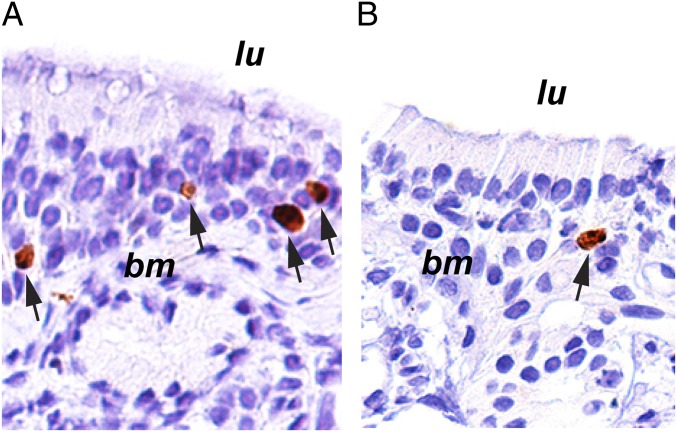
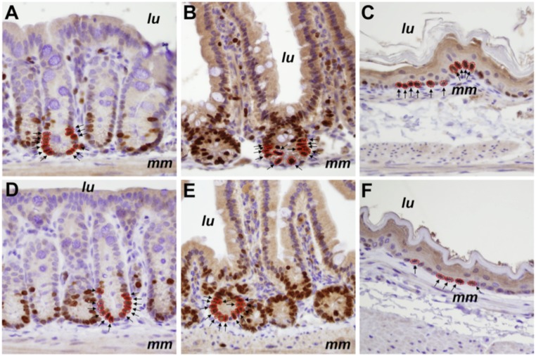
A new evaluation of previously published data suggested to us that the accumulation of mutations might slow, rather than increase, as individuals age. To explain this unexpected finding, we hypothesized that normal stem cell division rates might decrease as we age. To test this hypothesis, we evaluated cell division rates in the epithelium of human colonic, duodenal, esophageal, and posterior ethmoid sinonasal tissues. In all 4 tissues, there was a significant decrease in cell division rates with age. In contrast, cell division rates did not decrease in the colon of aged mice, and only small decreases were observed in their small intestine or esophagus. These results have important implications for understanding the relationship between normal stem cells, aging, and cancer. Moreover, they provide a plausible explanation for the enigmatic age-dependent deceleration in cancer incidence in very old humans but not in mice.

Keywords: aging; cancer; cell division; mutation rate.


Conflict of interest statement

The authors declare no conflict of interest.

Figures

Fig. 1.
Fig. 1.
Ki67 labeling of normal colon resections. Shown are examples of Ki67 labeling in a 26-y-old individual (A and B) compared with an 80-y-old individual (C and D). cr, properly oriented crypt; lu, location of the colonic lumen; mm, muscularis mucosae. The crypts outlined in A and C are magnified in B and D, respectively. Arrows indicate antibody-labeled nuclei, which are outlined in red. Images in A and C were taken at a magnification of 100×, while those in B and D were taken at a magnification of 400×.
Fig. 2.
Fig. 2.
Effects of aging on cell proliferation in the human colon. Differences in the proportion of Ki67-labeled cells (positions 1 to 8 from a crypt’s base for experiments 1A, 2, and 3; full crypt for experiment 1B) in colon (with first and third quartiles of the overall distribution) are illustrated.
Fig. 3.
Fig. 3.
Ki67 labeling of normal esophageal biopsies. Shown are examples of Ki67 labeling in a 25-y-old individual (A) compared with an 81-y-old individual (C). bm, basement membrane; lu, location of the esophageal lumen. The areas outlined in A and C are magnified in B and D, respectively. Arrows indicate antibody-labeled nuclei, which are outlined in red. Images in A and C were taken at a magnification of 100×, while those in B and D were taken at a magnification of 400×.
Fig. 4.
Fig. 4.
Effects of aging on cell proliferation in human esophagus. Differences in the proportion of Ki67-labeled cells (basal layer for experiments 1A, 2, and 3; full tissue for experiment 1B) in esophagus (with first and third quartiles of the overall distribution) are illustrated.
Fig. 5.
Fig. 5.
Ki67 labeling of normal duodenal biopsies. Shown are examples of Ki67 labeling in a 27-y-old individual (A) compared with an 84-y-old individual (C). lu, location of the duodenal lumen; mm, muscularis mucosae; vi, villus structures. The areas outlined in A and C are magnified in B and D, respectively. Arrows indicate antibody-labeled nuclei, which are outlined in red. Images in A and C were taken at a magnification of 100×, while images in B and D were taken at a magnification of 400×.
Fig. 6.
Fig. 6.
Effects of aging on cell proliferation in human duodenum and posterior ethmoid. Differences in the proportion of Ki67-labeled cells in duodenum (positions 1 to 8 from a crypt’s base) and in posterior ethmoid (with first and third quartiles of the overall distribution) are illustrated.
Fig. 7.
Fig. 7.
Ki67 labeling of normal posterior ethmoid sinonasal biopsies. Shown are examples of Ki67 labeling in a 32-y-old individual (A) compared with an 83-y-old individual (B). bm, basement membrane; lu, location of the sinus lumen. Arrows indicate antibody-labeled nuclei. Images were taken at a magnification of 20×.
Fig. 8.
Fig. 8.
Ki67 labeling of normal murine colon, small intestine, and esophagus. Shown are examples of Ki67 labeling in 1-mo-old (AC) compared with 25-mo-old (DF) male C57BL/6 mice. Images of colon (A and D), small intestine (B and E), and esophagus (C and F) were taken at a magnification of 400×. Both positively labeled (brown) and unlabeled (blue) nuclei can be seen in the photomicrographs. lu, location of the luminal surface; mm, muscularis mucosae. Examples of positive nuclei, either in crypts or in the basal layer, are indicated by arrows and are outlined in red.

References

    1. Araten D. J., et al. , A quantitative measurement of the human somatic mutation rate. Cancer Res. 65, 8111–8117 (2005). - PubMed
    1. DeMars R., Held K. R., The spontaneous azaguanine-resistant mutants of diploid human fibroblasts. Humangenetik 16, 87–110 (1972). - PubMed
    1. Drake J. W., Charlesworth B., Charlesworth D., Crow J. F., Rates of spontaneous mutation. Genetics 148, 1667–1686 (1998). - PMC - PubMed
    1. Tomasetti C., Vogelstein B., Parmigiani G., Half or more of the somatic mutations in cancers of self-renewing tissues originate prior to tumor initiation. Proc. Natl. Acad. Sci. U.S.A. 110, 1999–2004 (2013). - PMC - PubMed
    1. Welch J. S., et al. , The origin and evolution of mutations in acute myeloid leukemia. Cell 150, 264–278 (2012). - PMC - PubMed

Publication types

MeSH terms