. 2019 Oct 8;116(41):20428-20437.
doi: 10.1073/pnas.1901048116. Epub 2019 Sep 23.

Fibulin-4 exerts a dual role in LTBP-4L-mediated matrix assembly and function

Affiliations

Fibulin-4 exerts a dual role in LTBP-4L-mediated matrix assembly and function

Heena Kumra et al. Proc Natl Acad Sci U S A. .

Erratum in

Abstract

Elastogenesis is a hierarchical process by which cells form functional elastic fibers, providing elasticity and the ability to regulate growth factor bioavailability in tissues, including blood vessels, lung, and skin. This process requires accessory proteins, including fibulin-4 and -5, and latent TGF binding protein (LTBP)-4. Our data demonstrate mechanisms in elastogenesis, focusing on the interaction and functional interdependence between fibulin-4 and LTBP-4L and its impact on matrix deposition and function. We show that LTBP-4L is not secreted in the expected extended structure based on its domain composition, but instead adopts a compact conformation. Interaction with fibulin-4 surprisingly induced a conformational switch from the compact to an elongated LTBP-4L structure. This conversion was only induced by fibulin-4 multimers associated with increased avidity for LTBP-4L; fibulin-4 monomers were inactive. The fibulin-4-induced conformational change caused functional consequences in LTBP-4L in terms of binding to other elastogenic proteins, including fibronectin and fibrillin-1, and of LTBP-4L assembly. A transient exposure of LTBP-4L with fibulin-4 was sufficient to stably induce conformational and functional changes; a stable complex was not required. These data define fibulin-4 as a molecular extracellular chaperone for LTBP-4L. The altered LTBP-4L conformation also promoted elastogenesis, but only in the presence of fibulin-4, which is required to escort tropoelastin onto the extended LTBP-4L molecule. Altogether, this study provides a dual mechanism for fibulin-4 in 1) inducing a stable conformational and functional change in LTBP-4L, and 2) promoting deposition of tropoelastin onto the elongated LTBP-4L.

Keywords: LTBP-4; elastic fibers; fibrillin-1; fibronectin; fibulin-4.


Conflict of interest statement

The authors declare no conflict of interest.

Figures

Fig. 1.
Fig. 1.
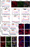
Fibulin-4 self-interacts via a central and a C-terminal region. (A) Schematic diagram of full-length fibulin-4 and the consecutive nonoverlapping Nterm-cbEGF1, cbEGF2-5, and cbEGF6-Cterm fragments (Left). SDS/PAGE of fibulin-4 fragments under reducing (+) and nonreducing (−) conditions (Right). The arrow points to high molecular mass species of cbEGF2-5 and cbEGF6-Cterm. (B) Mass size distributions of Nterm-cbEGF1, cbEGF2-5, and cbEGF6-Cterm determined by DLS. The x axis shows the particle hydrodynamic radius of the protein sample on a logarithmic scale, and the y axis the mass percentages of particles of a given hydrodynamic radius present in the sample. The peaks represent particle populations with radius sizes indicated on top. Note that the higher molecular mass protein bands in the SDS/PAGE for cbEGF6-Cterm and cbEGF2-5 (A, Right) correlate with peaks corresponding to larger sizes in DLS (B). (C and D) SPR sensorgrams of the interaction of the fibulin-4 fragments Nterm-cbEGF1, cbEGF2-5, and cbEGF6-Cterm (C), and of gel-filtrated full-length fibulin-4 monomers, dimers, and multimers (D) with full-length nongel-filtrated fibulin-4 immobilized on the chip. Note the binding of cbEGF2-5 and cbEGF6-Cterm in C and the particularly strong binding of multimers in D.
Fig. 2.
Fig. 2.
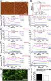
Fibulin-4 interacts with and assembles on fibronectin. (A) SPR sensorgram of the interaction of fibronectin (immobilized) with fibulin-4 (soluble). (B) Colocalization of fibronectin (red) with fibulin-4 (green) in the extracellular matrix produced by human skin fibroblasts 6 d after cell seeding, evident in the merged image (yellow). (C and D) SPR sensorgrams of the interaction of fibronectin (immobilized) with the soluble fibulin-4 fragments Nterm-cbEGF1, cbEGF2-5 and cbEGF6-Cterm (C), and with gel-filtrated full-length soluble fibulin-4 monomers, dimers, and multimers (D). Note that the binding sites for fibronectin are in the central and C-terminal region of fibulin-4, and that binding only occurs with multimers. (E) SPR sensorgrams of soluble Nterm-cbEGF1, cbEGF2-5, cbEGF6-Cterm, and full-length fibulin-4 as a control with the N-terminal half of fibrillin-1 (immobilized). Binding sites are located in the N-terminal cbEGF domain and the central region of fibulin-4. (F, Upper) Colocalization of fibulin-4 (red) with fibrillin-1 (green) in the matrix produced by human skin fibroblasts 6 d after cell seeding. (Lower) Fibrillin-1 (green) colocalizes with fibronectin (red) under identical conditions. (G) Immunofluorescence of fibulin-4 in the extracellular matrix of mouse skin fibroblasts either lacking Fbn-1 (Upper) or Fn (Lower) after addition of exogenous fibulin-4. Before analyzing the cells, 25 µg/mL purified fibulin-4 was added for 1 d. Note the absence of fibrillin-1 assembly in Fbn1−/− cells whereas fibulin-4 assembly was normal and comparable to Fbn1+/+ control cells. Both, fibronectin and fibulin-4 assembly was not detectable in the matrix produced by Fn−/− cells.
Fig. 3.
Fig. 3.
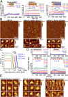
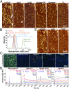
Conformational change in LTBP-4L induced by fibulin-4 multimers. (A) SPR sensorgrams of the interaction of Nterm-cbEGF1, cbEGF2-5 and cbEGF6-Cterm with immobilized LTBP-4L. (B) AFM height images showing LTBP-4L alone (Left), a 1:10 molar ratio of a LTBP-4L/fibulin-4 mixture in the presence of CaCl2 (Center), or 10 mM EDTA (Right) with higher magnification views of representative conformations of individual particles (Lower). Arrowheads indicate some elongated particles. (C) DLS mass size distribution of fibulin-4 alone, LTBP-4L alone, and a 1:10 molar ratio of a LTBP-4L/fibulin-4 mixture. The particle hydrodynamic radius is plotted logarithmically on the x axis and the mass percentages of particles on the y axis. Note the significant change in hydrodynamic radius upon mixing LTBP-4L with fibulin-4 as compared to fibulin-4 alone or LTBP-4L alone. ***P < 0.0001 (2-sample t test). (D) SPR sensorgrams of immobilized LTBP-4L with gel-filtrated full-length fibulin-4 monomers, dimers, multimers, and nongel-filtrated fibulin-4. Note that only fibulin-4 multimers and nongel-filtrated fibulin-4 (which contains multimers) interact with LTBP-4L. (E) AFM height images showing representative particles of mixtures of LTBP-4L with fibulin-4 monomers, dimers, and multimers at a 1:10 molar ratio (LTBP-4L:fibulin-4 monomer unit).
Fig. 4.
Fig. 4.
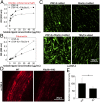
Functional change in LTBP-4L induced by fibulin-4. (A and B) Solid-phase binding assays of LTBP-4L and 1:10 molar ratio LTBP-4L/fibulin-4 mixture used as soluble ligand on coated fibrillin-1 N-terminal half (A) and fibronectin (B). A specific antiserum was used for detection of bound LTBP-4. Note that the LTBP-4L/fibulin-4 mixture (closed symbols) mediates a stronger binding to fibrillin-1 (A) and a reduced binding to fibronectin (B) as compared to LTBP-4L alone (open symbols). (C) Effect of fibulin-4–induced conformational change of LTBP-4L on LTBP-4 assembly in cell culture. For this, 10 µg/mL of LTBP-4L alone or 27 µg/mL of fibulin-4 alone or a mixture of 10 µg/mL of LTBP-4L and 27 µg/mL of fibulin-4 (molar ratio of 1:10) was added to human skin fibroblasts for 7 d prior to immunofluorescence analysis. TBS/2 mM CaCl2 was used as buffer control. Note the enhanced LTBP-4 assembly upon addition of the LTBP-4L/fibulin-4 mixture as compared to addition of the individual proteins. (D) LTBP-4 staining in wild-type (WT) and fibulin-4 knockout (KO) aorta harvested at embryonic day 17.5. (E) Quantification of the staining in (D). *P < 0.05 (2-sample t test). Note the reduced LTBP-4 staining, indicating reduced assembly/deposition in the absence of fibulin-4.
Fig. 5.
Fig. 5.
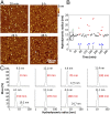
Dynamics of fibulin-4 induced conformational change in LTBP-4L. (A) AFM height images showing a time course analysis of a LTBP-4L/fibulin-4 mixture (1:10 molar ratio) at endpoints 20 min, 6 h, 24 h, and 48 h after mixing. Note that the fibulin-4–induced conformational change of LTBP-4L, which is observed as early as 20 min (also evident from Fig. 3B), did not reverse within the analysis time. (B) Real-time DLS mass distribution analysis of a LTBP-4L/fibulin-4 mixture (1:10 molar ratio) within 0 to 300 min. Black symbols represent data points where a single peak was observed, red and blue symbols represent the larger and smaller size data points, respectively, when 2 peaks appeared. At each time point there was either 1 single peak (black symbol) or 2 peaks (red and blue symbol), but never 3 peaks. The boxed area highlights the time span in which extension of LTBP-4L becomes fully induced. The green arrow points to a 20-min time point after which the extension was stable. The white arrows show a periodic pattern of dissociation. (C) Selected mass distribution plots obtained from the real-time DLS analysis (plotted in B), demonstrating events of association and conformational changes between LTBP-4L and fibulin-4 (0 to 20 min), and events of periodic dissociation and association between the extended LTBP-4L and fibulin-4. The x axis shows on a logarithmic scale the particle hydrodynamic radius of the protein sample in nanometers, and the y axis represents the mass percentages of particles with a given hydrodynamic radius in the sample.
Fig. 6.
Fig. 6.
A transient interaction with fibulin-4 is sufficient to induce conformational and functional change in LTBP-4L. (A) AFM height images showing LTBP-4L and fibulin-4 primed LTBP-4L. Arrowheads indicate some elongated molecules. (B) DLS mass distribution of fibulin-4–primed LTBP-4L (red curve) overlaid the profiles for fibulin-4, LTBP-4L, and of a 1:10 molar ratio LTBP-4L/fibulin-4 mixture from Fig. 3C (gray curves). Note the similarity in the hydrodynamic radius between fibulin-4–primed LTBP-4L and the LTBP-4L/fibulin-4 mixture, and the significant differences compared to fibulin-4 alone or LTBP-4L alone. The x axis shows the particle hydrodynamic radius on a logarithmic scale and the y axis the mass percentages of particles. ***P < 0.0001; ns indicates a nonsignificant P value (2-sample t test). (CF) SPR sensorgrams of LTBP-4L (nonprimed; Left) or fibulin-4–primed LTBP-4L (Right) binding to fibrillin-1 N-terminal half (C), fibronectin (D), fibulin-4 (E), and to itself (LTBP-4L to LTBP-4L, and fibulin-4–primed LTBP-4L to fibulin-4–primed LTBP-4L) (F). Note the changes in binding affinities. (G) Comparison of LTBP-4L self-assembly by immunofluorescence between fibulin-4–primed LTBP-4L (10 µg/mL) and a mixture of LTBP-4L and fibulin-4 (10 µg/mL LTBP-4L and 27 µg/mL fibulin-4 equivalent to a 1:10 molar ratio) after addition to human skin fibroblasts. (H) Quantification of the total fiber length in G. Note that both conditions similarly promote LTBP-4L assembly.
Fig. 7.
Fig. 7.
Role of fibulin-4 and LTBP-4L in tropoelastin aggregation and binding. (A) AFM height images of tropoelastin (TE) together with either the LTBP-4L/fibulin-4 mixture (L4/F4/TE), the fibulin-4–primed LTBP-4L (L4-primed/TE), or fibulin-4 (F4/TE). As controls, the LTBP-4L/fibulin-4 mixture (L4/F4), the fibulin-4 primed LTBP-4L (L4-primed), or TE alone are shown. (B) DLS mass distribution of TE alone or mixed with either fibulin-4 (F4/TE), LTBP-4L (L4/TE), fibulin-4–primed LTBP-4L (L4-primed/TE) or the LTBP-4L/fibulin-4 mixture (L4/F4/TE). The x axis represents the particle hydrodynamic radius on a logarithmic scale, and the y axis the particle mass percentages. Note the shift in hydrodynamic radius to large size (B) and the large assembly structures formed (A) only in the presence of LTBP-4L, fibulin-4, and tropoelastin together. (C) Elastin assembly in the extracellular matrix of human skin fibroblasts supplemented with either a mixture of 10 µg/mL LTBP-4L and 27 µg/mL fibulin-4 (1:10 molar ratio), or 10 µg/mL fibulin-4 primed LTBP-4L, or 10 µg/mL LTBP-4L alone, or 27 µg/mL fibulin-4 alone. TBS/2 mM CaCl2 was used as buffer control. (D) SPR binding assays with soluble Nterm-cbEGF1, cbEGF2-5, and cbEGF6-Cterm fibulin-4 fragments and immobilized tropoelastin. Note that the binding sites are located in the center and the C-terminal region of fibulin-4. (E) AFM height images of tropoelastin (TE) together with either a LTBP-4L/fibulin-5 mixture (L4/F5/TE) or fibulin-5 (F5/TE). As control, the LTBP-4L/fibulin-5 mixture (L4/F5) is shown.
Fig. 8.
Fig. 8.
Schematic representation of the proposed elastogenic model highlighting the role of fibulin-4 and LTBP-4L. Details are described in the text.

References

    1. Papke C. L., Yanagisawa H., Fibulin-4 and fibulin-5 in elastogenesis and beyond: Insights from mouse and human studies. Matrix Biol. 37, 142–149 (2014). - PMC - PubMed
    1. Kinsey R., et al. , Fibrillin-1 microfibril deposition is dependent on fibronectin assembly. J. Cell Sci. 121, 2696–2704 (2008). - PubMed
    1. Sabatier L., et al. , Fibrillin assembly requires fibronectin. Mol. Biol. Cell 20, 846–858 (2009). - PMC - PubMed
    1. Kantola A. K., Keski-Oja J., Koli K., Fibronectin and heparin binding domains of latent TGF-beta binding protein (LTBP)-4 mediate matrix targeting and cell adhesion. Exp. Cell Res. 314, 2488–2500 (2008). - PubMed
    1. Ono R. N., et al. , Latent transforming growth factor beta-binding proteins and fibulins compete for fibrillin-1 and exhibit exquisite specificities in binding sites. J. Biol. Chem. 284, 16872–16881 (2009). - PMC - PubMed

LinkOut - more resources