Review
. 2019 Sep 23;10(10):738.
doi: 10.3390/genes10100738.

Molecular Therapies for Choroideremia

Affiliations
Review

Molecular Therapies for Choroideremia

Jasmina Cehajic Kapetanovic et al. Genes (Basel). .

Abstract

Advances in molecular research have culminated in the development of novel gene-based therapies for inherited retinal diseases. We have recently witnessed several groundbreaking clinical studies that ultimately led to approval of Luxturna, the first gene therapy for an inherited retinal disease. In parallel, international research community has been engaged in conducting gene therapy trials for another more common inherited retinal disease known as choroideremia and with phase III clinical trials now underway, approval of this therapy is poised to follow suit. This chapter discusses new insights into clinical phenotyping and molecular genetic testing in choroideremia with review of molecular mechanisms implicated in its pathogenesis. We provide an update on current gene therapy trials and discuss potential inclusion of female carries in future clinical studies. Alternative molecular therapies are discussed including suitability of CRISPR gene editing, small molecule nonsense suppression therapy and vision restoration strategies in late stage choroideremia.

Keywords: REP1; choroideremia; gene therapy; inherited retinal disease; treatment.


Conflict of interest statement

REM is a founder and receives grant funding from Nightstar Therapeutics (now Biogen Inc.). REM and ARB are consultants to Nightstar Therapeutics and REM is a consultant to Spark Therapeutics. These companies did not have any input into the work presented.

Figures

Figure 1
Figure 1
Retinal imaging in choroideremia. Widefield optomaps, Optos, Dumfernline, UK (A,B) and Heidelberg Spectralis imaging, Heidelberg, Germany (CF) showing choroideremia phenotype in an affected male. Colour fundus photographs (C,D) show extensive retinal degeneration with choroidal atrophy and visualisation of underlying pale sclera. Fundus autofluorescence (E,F) shows typical patterns of sharply demarcated areas of remaining tissue (hyperfluorescent) against atrophic retina (hypofluorescent background). Mesopic microperimetry, MAIA CenterVue SpA, Padova, Italy (G,H) measures central retinal sensitivity that closely maps areas of residual retina as seen on autofluorescence. Sensitivity maps are shown with corresponding histograms of threshold frequencies. Spectral domain optical coherence tomography, Heidelberg, Germany (I,J) shows retinal structure in cross-section with distribution of ellipsoid zone (yellow line) and preserved inner retinal layers.
Figure 2
Figure 2
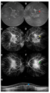
Retinal imaging in a choroideremia patient showing an area of scaring from an old choroidal neovascular membrane in the left eye. Fundus autofluorescence (A, B), fluorescein angiography (C, D), indocyanine green angiography (E, F) and spectral domain optical coherence tomography (G) with arrows marking the old scar. Imaging was performed with Heidelberg Spectralis, Heidelberg, Germany.
Figure 3
Figure 3
Retinal imaging in two female choroideremia carriers. Phenotype of an asymptomatic mild carrier with Snellen visual acuity of 6/5 in both eyes is shown from (AF) and a carrier with a ‘geographic-pattern’ phenotype and reduced visual acuity of 6/7.5 in the right eye and 6/12 in the left eye is shown from (GL). Fundus autofluorescence showing very early signs of fine ‘salt and pepper’ mottling (A,B) compared with coarse mottling and atrophic patches resembling geographic patterns (G,H). Mesopic microperimetry, MAIA CenterVue SpA, Padova, Italy showing sensitivity maps with corresponding histograms of threshold frequencies. Near-normal central retinal sensitivity is found in mild, asymptomatic carriers (C,D) compared to reduced retinal sensitivity in affected carriers especially in the left eye of the above case (I,J). OCT imaging is clinically insignificant in mild, asymptomatic carriers (E, F) whereas some disruption of retinal pigment epithelium (RPE) and ellipsoid zone is observed in the affected carrier, particularly in the left eye (K,L).

References

    1. Cremers F.P., van de Pol D.J., van Kerkhoff L.P., Wieringa B., Ropers H.H. Cloning of a gene that is rearranged in patients with choroideraemia. Nature. 1990;347:674–677. doi: 10.1038/347674a0. - DOI - PubMed
    1. Seabra M.C., Brown M.S., Goldstein J.L. Retinal degeneration in choroideremia: Deficiency of rab geranylgeranyl transferase. Science. 1993;259:377–381. doi: 10.1126/science.8380507. - DOI - PubMed
    1. Van den Hurk J.A., Schwartz M., van Bokhoven H., Van de Pol T.J.R., Bogerd L., Pinckers A.J.L.G., Bleeker-Wagemakers E.M., Pawlowitzki I.H., Rüther K., Ropers H.H., et al. Molecular basis of choroideremia (CHM): Mutations involving the Rab escort protein-1 (REP-1) gene. Hum. Mutat. 1997;9:110–117. doi: 10.1002/(SICI)1098-1004(1997)9:2<110::AID-HUMU2>3.0.CO;2-D. - DOI - PubMed
    1. Tolmachova T., Anders R., Abrink M., Bugeon L., Dallman M.J., Futter C.E., Ramalho J.S., Tonagel F., Tanimoto N., Seeliger M.W., et al. Independent degeneration of photoreceptors and retinal pigment epithelium in conditional knockout mouse models of choroideremia. J. Clin. Investig. 2006;116:386–394. doi: 10.1172/JCI26617. - DOI - PMC - PubMed
    1. Patrício M.I., Barnard A.R., Xue K., MacLaren R.E. Choroideremia: Molecular mechanisms and development of AAV gene therapy. Expert Opin. Biol. Ther. 2018;18:807–820. doi: 10.1080/14712598.2018.1484448. - DOI - PubMed

Publication types