Review
. 2017 Oct 25;6(4):16.
doi: 10.3390/antib6040016.

Antibodies and Derivatives Targeting DR4 and DR5 for Cancer Therapy

Affiliations
Review

Antibodies and Derivatives Targeting DR4 and DR5 for Cancer Therapy

Agathe Dubuisson et al. Antibodies (Basel). .

Abstract

Developing therapeutics that induce apoptosis in cancer cells has become an increasingly attractive approach for the past 30 years. The discovery of tumor necrosis factor (TNF) superfamily members and more specifically TNF-related apoptosis-inducing ligand (TRAIL), the only cytokine of the family capable of eradicating selectively cancer cells, led to the development of numerous TRAIL derivatives targeting death receptor 4 (DR4) and death receptor 5 (DR5) for cancer therapy. With a few exceptions, preliminary attempts to use recombinant TRAIL, agonistic antibodies, or derivatives to target TRAIL agonist receptors in the clinic have been fairly disappointing. Nonetheless, a tremendous effort, worldwide, is being put into the development of novel strategic options to target TRAIL receptors. Antibodies and derivatives allow for the design of novel and efficient agonists. We summarize and discuss here the advantages and drawbacks of the soar of TRAIL therapeutics, from the first developments to the next generation of agonistic products, with a particular insight on new concepts.

Keywords: TRAIL; antibody; antibody drug conjugate; apoptosis; bi-specific; cancer therapy; chimeric antigen receptor; death-receptor targeting; ligand; scFv.


Conflict of interest statement

The authors declare no conflict of interest.

Figures

Figure 1
Figure 1
Schematic representation of TRAIL receptors. TRAIL-R1/death receptor 4 (DR4), TRAIL-R2/death receptor 5 (DR5), act as agonistic receptors, owing to their intracellular death domain (DD), through which they transmit an apoptotic signal. The other two membrane receptors, TRAIL-R3/decoy receptor 1 (DcR1) and TRAIL-R4/decoy receptor 2 (DcR2), act as antagonistic/regulatory receptors, due to their lack of functional DD. Osteoprotegerin (OPG), contains a DD, but is unable to trigger apoptosis as it lacks a transmembrane domain (TM), and is therefore a soluble receptor. DR4 and DR5 have been found to be N- and O-glycosylated, respectively [22,23,24]. DcR1, DcR2 and OPG, alike, as most members of the TNF superfamily [37], contain putative glycosylation sites, which are depicted as blue stars or red triangle. The size of these receptors is shown on the right-hand side.
Figure 2
Figure 2
Schematic representation of the amino acid composition of TRAIL agonist receptors and crystallographic structure of the TRAIL/receptor complexes. (A) Alignment of DR4, DR5a, and DR5b. Sequence identity is shown. Orange and red patches correspond to shared surface residues; (B) Surface representation of trimeric TRAIL with DR4 and DR5 from the crystallographic structures 1D4V [42] and 5CIR [43], respectively. Top and side views are shown. Shared surface residues between DR4 and DR5 are shown by the two patches (orange and red). Buried or non-shared residues are shown in grey. In the trimeric representations, TRAIL monomers are shown as cornflower, medium, and dark blue. DR4 or DR5 monomers are shown in grey. DR4 and DR5 N-glycosylation and O-glycosylation sites are depicted with the following symbols # and *, respectively.
Figure 3
Figure 3
Simplified schematic representation of the main events regulating TRAIL-induced apoptosis. DR4 and DR5 agonistic receptors are able to recruit FADD and caspase-8 upon TRAIL stimulation, leading to apoptosis (see text for detail). In the presence of antagonist receptors, TRAIL-induced cell death is restrained, either due to competition for TRAIL binding (DcR1 and OPG) or steric hindrance, leading to reduced caspase-8 activation (DcR2, see Merino et al., 2006 [47], and Shirley [41]). Within the cytoplasm, the inhibitor c-FLIP, as well as Bcl-2 family members, can also restrain caspase activation and apoptosis, leading to cell resistance to TRAIL-induced cell death [48].
Figure 4
Figure 4
Schematic representation of the main TRAIL recombinant proteins assessed in clinical trials or used in laboratories to induce apoptosis through DR4 and/or DR5. Dulanermin (TRAIL aa114-281) [65,66], circularly permuted TRAIL (CPT, TRAIL aa135-281-linker-TRAIL aa122-135) [67], Flag-TRAIL [68], TNC-TRAIL [61], 6xHis-TRAIL [55], HSA-TRAIL [69], Leucine Zipper and Isoleucine Zipper-TRAIL [15], Trimer-TRAIL [70], Fc-TRAIL [63], Fc-sc-TRAIL [71], and AGP350 [40].
Figure 5
Figure 5
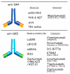
Schematic representation of the DR4 and DR5 moAb. Anti-DR4 developed in preclinical and clinical trials are shown in the upper panel. m921/922, 4H6 & 4G7, AY4, and TR1-mAbs are mouse-Igs tested in preclinical trials. Mapatumumab is a humanized-Ig assessed in clinical trials. The lower part of the figure shows the anti-DR5 developed as mouse or chimeric-Igs, which are LaDR5, LBY135, mDRA, WD1, and Zaptuximab, as well as its humanized form, Zaptuzumab. Conatumumab, Drozitumab, Tigatuzumab, KMTR2, Lexatumumab, and more recently, DS-8273a, are also humanized antibodies targeting DR5. All of them have been tested in clinical trials.
Figure 6
Figure 6
Schematic representation of DR4 or DR5 multivalent agonists. Represented on the upper left is the pentavalent IgM form of the newly described anti-DR4 antibodies [161]. The upper right shows tetravalent scFvs targeting DR5 [153] and TAS266 nanobody [154]. In the lower part, primary structures of DR5-peptidomimetics as monomer, dimer, and trimer are represented [152].
Figure 7
Figure 7
Schematic representation of scFv-TRAIL and non-scFv-TRAIL constructs. Upper left: presentation of different cancer (DsG3 [162], EGFR [149,150], EGP2 [152], Kv10.1 [153], MCSP [155], or MRP3 [156]) and immune (CD3 [163], CD7 [164], CD19 [165,166], CD20 [167], CD33 [168], CD40 [169], CD47 [170], CD70 [171], CLL1 [172], or PD-L1 [173]) antigens targeted by scFv-TRAIL constructs. Upper right: single-chain TRAIL-bispecific derivatives targeting EGFR [40,71], ErbB2 [151], and mesothelin [154]. Lower part: non-scFv-TRAIL bi- and trispecific derivatives using nanobody (ENb:TRAIL), affibody, or fusion proteins, to target cancer and immune antigens.
Figure 8
Figure 8
Schematic representation of unconventional and BsAbs harboring selectivity for DR4 and/or DR5. Fibronectin type III domain of tenascin C-based formats recognizing DR5, are presented in the upper left, covalently linked to Igs, Fc or as single-chains. Amino acids highlighted in red/orange and pale blue correspond to TN3 variable loops (DE, FG and BC) which can be mutated without changing its globular structure. These correspond to aa that allow binding to DR5. Amino acids shown in beige, depict TN3 amino acids that need to be preserved to maintain the globular structure of the recombinant protein. Upper right panel illustrates Kringle domain (KD)-based formats fused to the Fc. Specific KDs are numbered 413 to 548. Affinity binding for DR4 and/or DR5 is indicated. Lower left panel depicts anti-DR4 and/or DR5–derivatives targeting TRA-8, LTβR, FAP, MCSP, or hyaluronate. BsAbs targeting both DR4 and DR5 are represented in the lower right.
Figure 9
Figure 9
Schematic representation of chimeric antigen receptor (CAR). Here, the CAR construct is presented, containing a scFv extracellular domain targeting DR4, a CD8 hinge and a CD3 intracellular domain mediating T-cell activation. This compound is capable of activating both DR4-mediated apoptosis and tumor specific T-cell cytotoxicity.
Figure 10
Figure 10
TRAIL ADC-like derivatives. From the left to the right : ADC-like TRAIL build up with Smac-derived peptide (AD-053.2) an intracellular pro-apoptotic agent [225]; Melittin ADC-like, an antibacterial highly cytotoxic harbored on trimeric TRAIL and linked with FLAG [226,227]; MMAE–TRAIL, TRAIL trimeric molecule linked to the monomethyl auristatin E, an antimitotic agent [222,223,224].

References

    1. Kerr J.F., Wyllie A.H., Currie A.R. Apoptosis: A basic biological phenomenon with wide-ranging implications in tissue kinetics. Br. J. Cancer. 1972;26:239–257. doi: 10.1038/bjc.1972.33. - DOI - PMC - PubMed
    1. DeVita V.T., Jr., Chu E. A history of cancer chemotherapy. Cancer Res. 2008;68:8643–8653. doi: 10.1158/0008-5472.CAN-07-6611. - DOI - PubMed
    1. Carswell E.A., Old L.J., Kassel R.L., Green S., Fiore N., Williamson B. An endotoxin-induced serum factor that causes necrosis of tumors. Proc. Natl. Acad. Sci. USA. 1975;72:3666–3670. doi: 10.1073/pnas.72.9.3666. - DOI - PMC - PubMed
    1. Lejeune F.J., Ruegg C., Lienard D. Clinical applications of TNF-alpha in cancer. Curr. Opin. Immunol. 1998;10:573–580. doi: 10.1016/S0952-7915(98)80226-4. - DOI - PubMed
    1. Trauth B.C., Klas C., Peters A.M., Matzku S., Möller P., Falk W., Debatin K.M., Krammer P.H. Monoclonal antibody-mediated tumor regression by induction of apoptosis. Science. 1989;245:301–305. doi: 10.1126/science.2787530. - DOI - PubMed

LinkOut - more resources