. 2019 Sep 23;9(1):13694.
doi: 10.1038/s41598-019-50114-2.

Novel mitochondrial complex I-inhibiting peptides restrain NADH dehydrogenase activity

Affiliations

Novel mitochondrial complex I-inhibiting peptides restrain NADH dehydrogenase activity

Yao-Peng Xue et al. Sci Rep. .

Abstract

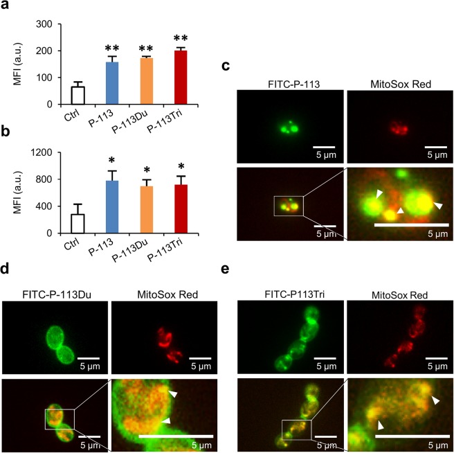
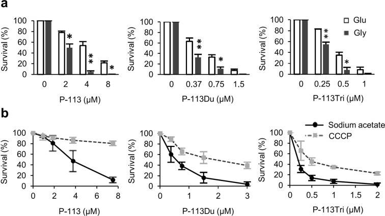
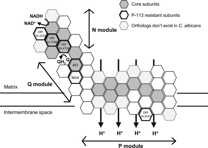
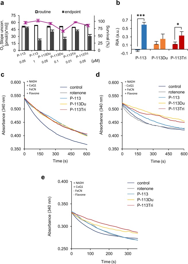
The emergence of drug-resistant fungal pathogens is becoming increasingly serious due to overuse of antifungals. Antimicrobial peptides have potent activity against a broad spectrum of pathogens, including fungi, and are considered a potential new class of antifungals. In this study, we examined the activities of the newly designed peptides P-113Du and P-113Tri, together with their parental peptide P-113, against the human fungal pathogen Candida albicans. The results showed that these peptides inhibit mitochondrial complex I, specifically NADH dehydrogenase, of the electron transport chain. Moreover, P-113Du and P-113Tri also block alternative NADH dehydrogenases. Currently, most inhibitors of the mitochondrial complex I are small molecules or artificially-designed antibodies. Here, we demonstrated novel functions of antimicrobial peptides in inhibiting the mitochondrial complex I of C. albicans, providing insight in the development of new antifungal agents.


Conflict of interest statement

The authors declare no competing interests.

Figures

Figure 1
Figure 1
P-113, P-113Du and P-113Tri target mitochondria. The intracellular ROS levels were measured using the fluorescent dye H2DCFDA (a) and MitoSOX Red (b) after the cells were treated with a sublethal dose of the AMPs at 37 °C for 1 h. The results are represented as the mean ± SD of three independent experiments. P-values are derived from the cells treated with AMPs compared to the control cells without treatment. *P < 0.05; **P < 0.01. Fluorescent images of C. albicans with FITC (green)-labeled P-113 (c), P-113Du (d), and P-113Tri (e). Mitochondria were stained with MitoSox Red (red). The white triangles point to the colocalization of AMPs and mitochondria. Images of cells without AMP treatment are provided in Supplementary Fig. 2.
Figure 2
Figure 2
The AMPs affect cellular respiration. (a) Cells were grown in glycerol (Gly, 2% w/v) or glucose (Glu, 2% w/v) for 5 h, followed by the addition of AMPs and further incubation at 37 °C for 1 h. *P < 0.05; **P < 0.01 for cells grown in Glu vs. Gly. (b) Cells were grown at 37 °C for 1 h in the presence of AMP alone (dissolved in sodium acetate) or AMP + CCCP. Cell viability was determined by measuring colony-forming units that were normalized to the control without AMP treatment (as 100%). The results are represented as the mean ± SD of three independent experiments.
Figure 3
Figure 3
C. albicans mitochondrial complex I subunits are demonstrated. The possible distribution of core subunits in mitochondrial complex I is shown in gray blocks. Mutants resistant to P-113 are also indicated (outlined in bold). ORF19.7590, ORF19.1710 (Ali1) and ORF19.4758 appear to be core subunits that are conserved from bacteria to mammalian cells. ORF19.1625, ORF19.2570 (Mci4) and ORF19.5547 appear to be accessary subunits of mitochondrial complex I. These proteins, which are possibly targeted by P-113, are mostly components of the hydrophilic domain of complex I. This diagram is drawn based on the structural models of mitochondrial complex I from She et al..
Figure 4
Figure 4
The AMPs effectively inhibit respiration and NADH dehydrogenases. (a) The oxygen consumption rates and survival rates (purple line) of cells treated with AMPs are shown. The survival rates were determined by recording the number of colony-forming units from the cells used for the oxygen consumption assays. The white bar represents the routine respiration rate of the cells and the black bar represents the endpoint of the respiration rate after AMP addition. The results are presented as the mean ± SD of three independent experiments. (b) The relative inhibitory activity (RIA) was determined by normalization of the inhibitory rate to the survival rate of the AMP-treated cells. The AMP concentrations from left to right are P-113 (0.05 μM), P-113 (1 μM), P-113Du (0.05 μM), P-113Du (0.1 μM), P-113Tri (0.01 μM), P-113Tri (0.05 μM). (c) Mitochondria were isolated from C. albicans and treated with rotenone or various AMPs in the presence of flavone with CoQ1 as the electron acceptor. NADH consumption was determined by measuring the absorbance at 340 nm. (d) The inhibitory effect of AMPs on NADH oxidization by complex I was assayed using ferricyanide instead of CoQ1 as the electron acceptor. (e) The effects of AMPs on alternative NADH dehydrogenases using an assay similar to (c) but without the addition of flavone. For (c) to (e), all experiments were repeated independently at least three times and the representative plots from three experiments with similar results are shown.
Figure 5
Figure 5
ROS induction is associated with the candidacidal activity of AMPs. (a) The levels of malondialdehyde, a lipid peroxidation marker, were measured at 530 nm in cells treated with or without AMPs for 2 h. The MDA content was calculated and expressed as 10−3 μM/mg protein. *P < 0.05; **P < 0.01 for cells with AMP treatment vs. the control (without AMP treatment). (b) The ROS scavenger ascorbic acid reduces cellular susceptibility to AMPs. C. albicans cells were treated with AMPs, incubated at 37 °C for 1 h, plated on YPD agar and the colony-forming units (cfu) were counted. The number of cfus of the AMP-treated cells were normalized to the control cells without treatment. (c) The intracellular ATP content of AMP-treated C. albicans cells was measured using a luciferase-based assay. Luminescence was measured at 560 nm. ATP content is shown as (μM)/mg protein. The results are represented as the mean ± SD of three independent experiments.

References

    1. Bahar AA, Ren D. Antimicrobial peptides. Pharmaceuticals (Basel) 2013;6:1543–1575. doi: 10.3390/ph6121543. - DOI - PMC - PubMed
    1. Wang G. Human antimicrobial peptides and proteins. Pharmaceuticals (Basel) 2014;7:545–594. doi: 10.3390/ph7050545. - DOI - PMC - PubMed
    1. Ng SMS, et al. Antifungal peptides: a potential new class of antifungals for treating vulvovaginal candidiasis caused by fluconazole‐resistant Candida albicans. Journal of Peptide Science. 2017;23:215–221. doi: 10.1002/psc.2970. - DOI - PubMed
    1. Rothstein DM, et al. Anticandida activity is retained in P-113, a 12-amino-acid fragment of histatin 5. Antimicrob Agents Chemother. 2001;45:1367–1373. doi: 10.1128/AAC.45.5.1367-1373.2001. - DOI - PMC - PubMed
    1. Driscoll J, et al. Candidacidal activity of human salivary histatin recombinant variants produced by site-directed mutagenesis. Gene. 1996;177:29–34. doi: 10.1016/0378-1119(96)00265-X. - DOI - PubMed

Publication types

MeSH terms