. 2019 Oct;15(10):975-982.
doi: 10.1038/s41589-019-0370-y. Epub 2019 Sep 23.

The morphogen Sonic hedgehog inhibits its receptor Patched by a pincer grasp mechanism

Affiliations

The morphogen Sonic hedgehog inhibits its receptor Patched by a pincer grasp mechanism

Amalie F Rudolf et al. Nat Chem Biol. 2019 Oct.

Abstract

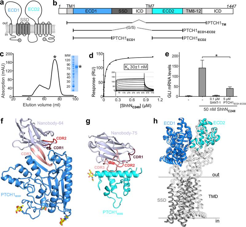
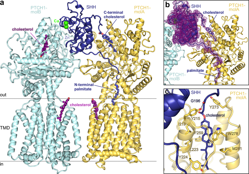
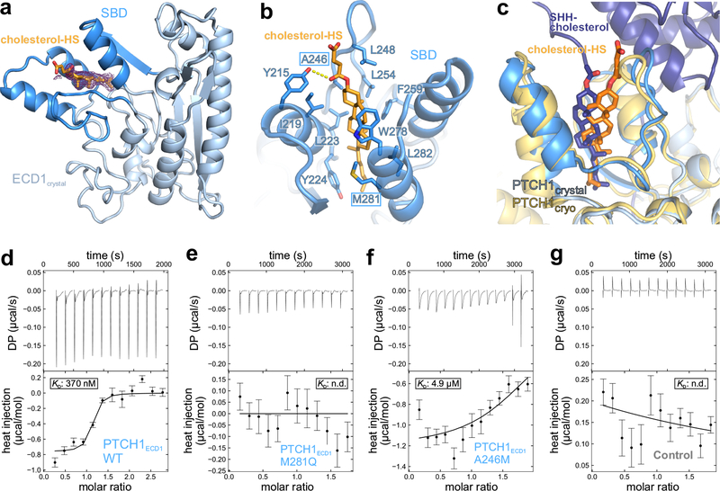
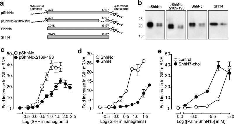
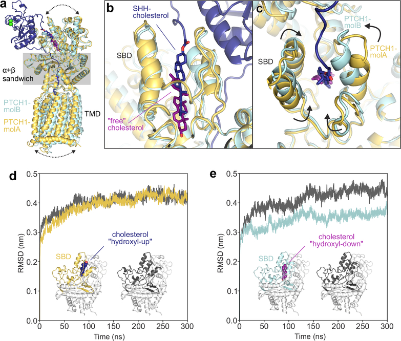
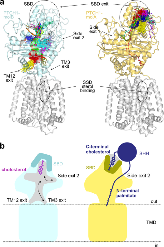
Hedgehog (HH) ligands, classical morphogens that pattern embryonic tissues in all animals, are covalently coupled to two lipids-a palmitoyl group at the N terminus and a cholesteroyl group at the C terminus. While the palmitoyl group binds and inactivates Patched 1 (PTCH1), the main receptor for HH ligands, the function of the cholesterol modification has remained mysterious. Using structural and biochemical studies, along with reassessment of previous cryo-electron microscopy structures, we find that the C-terminal cholesterol attached to Sonic hedgehog (Shh) binds the first extracellular domain of PTCH1 and promotes its inactivation, thus triggering HH signaling. Molecular dynamics simulations show that this interaction leads to the closure of a tunnel through PTCH1 that serves as the putative conduit for sterol transport. Thus, Shh inactivates PTCH1 by grasping its extracellular domain with two lipidic pincers, the N-terminal palmitate and the C-terminal cholesterol, which are both inserted into the PTCH1 protein core.


Conflict of interest statement

Competing interests

The authors declare no competing interests.

Figures

Figure 1 |
Figure 1 |. Structural and function characterization of PTCH1-nanobody interactions.
a, The pseudo-symmetric domain architecture of PTCH1: two 6-TM segments with extracellular domains (ECD1 and ECD2) interposed between the first two TM helices of each segment. The SSD, composed of TM helices 2–6, is marked in gray. b, Composition of the various protein constructs used in this study. c-e, Characterization of PTCH1ECD1-ECD2 used as an antigen to immunize Llamas. c, Typical SEC purification and corresponding SDS-PAGE of pooled fractions of PTCH1ECD1-ECD2. d, SPR equilibrium binding experiment between PTCH1ECD1-ECD2 (ligand) and non-lipidated ShhNC24II (analyte). This experiment was independently repeated 3 times with similar results. e, SHH signalling assay in mouse NIH-3T3 cells. Normalized Gli1 mRNA expression was used to assess SHH signalling activity by RT-qPCR after stimulation with purified ShhNC24II. Error bars denote SEM of 3 independent experiments. Statistical value is p=0.0387 determined by ordinary one-way ANOVA with Turkey’s multiple comparisons test. Sant-1 is a HH signalling inhibitor acting downstream of PTCH1. PTCH1ECD1-ECD2 acts as a ligand trap to inhibit Hh signalling. f-g, Cartoon representations of the high-resolution crystal structures of the PTCH1ECD1-NB64 (f) and PTCH1ECD2-NB75 (g) complexes. The complementary determining regions (CDRs) of the nanobodies are highlighted and N-linked glycans are shown in atomic colouring (carbon: yellow, oxygen: red, nitrogen: blue). h, Superposition of the PTCH1ECD1 and PTCH1ECD2 crystal structures on the previously determined cryo-EM PTCH1TM structure (PDB 6E1H).
Figure 2 |
Figure 2 |. Structure of the PTCH1-pShhNc complex.
a, Rebuilt and improved model of the complex between one molecule of pShhNc and two molecules of PTCH1 (molA and molB). The high-affinity protein-protein interface between PTCH1-molB (cyan) and pShhNc is organized by the highly conserved calcium (green)- and zinc (grey)-binding sites of pShhNc (dark blue). The interface with PTCH1-molB (yellow) is composed of the cholesteryl- and palmitoyl- appendages of pShhNc that simultaneously penetrate the ECD. b, The previously deposited cryo-EM map of the SHH-PTCH1 complex (EMD-8955) mapped onto our improved model. Extra density is clearly discernible stretching from the C-terminus of pShhNc into ECD1 of PTCH1. c, Close-up view of the SHH-cholesterol molecule bound in the sterol-binding domain (SBD) of PTCH1, with residues in contact with cholesterol depicted in stick representation.
Figure 3 |
Figure 3 |. Structural and biophysical characterisation of the PTCH1 ECD-cholesterol complex.
a, Crystal structure of the PTCH1ECD1-cholesterol-hemisuccinate (cholesterol-HS) complex, with the 3-helix SBD coloured dark blue. The initial 2Fo-Fc map at 1.0 σ and 1.9 Å resolution before inclusion of cholesterol-HS is shown in wire representation. b, Close-up view of the sterol-binding site with cholesterol-HS depicted in orange sticks. c, Superposition of the PTCH1ECD1-cholesterol-HS crystal structure on the improved PTCH1-pShhNc cryo-EM structure. The entrance of the sterol-binding pocket is slightly rearranged, likely due to different steric constraints of the hemisuccinate group compared to the glycine ester linkage found in pShhNc. d-g, Raw ITC data (upper panel) and binding isotherms (lower panel) for titration of PEG-cholesterol into solutions containing PTCH1ECD1-WT (d), PTCH1ECD1-M281Q (e) and PTCH1ECD1-A246M (f). PEG200, unconjugated to cholesterol, was titrated into a PTCH1ECD1-WT solution as a control (g). Curves show the A+B ⇌AB binding model that was used for local fitting to the data. The Kd is shown where it could be calculated. Bars shown on the isotherm reflect errors associated with integration of the injection peaks in the corresponding thermograms (68% confidence around extrapolation of pre-and post-injection baselines).
Figure 4 |
Figure 4 |. The cholesterol attached to pShhNc inactivates PTCH1.
a, Schematic of the various SHH proteins used in signalling assays. b, Two different amounts of each SHH variant from (h) were analyzed by immunoblotting. Full gels can be found on Supplementary Figure 7. c and d, Fold-increase in Gli1 mRNA (relative to the no SHH added condition) was used as a metric for signalling strength after treatment of NIH/3T3 cells with various concentrations of the indicated ligands. e, HH signalling strength at increasing concentrations of the N-terminal palmitoylated peptide (Palm-ShhN15) in the absence or presence (100 μM) of a cholesteroylated C-terminal peptide (ShhN7-chol). In c-e, the mean is depicted and error bars reflect standard deviation (n=4). Each experiment in c-e was repeated at least 3 times.
Figure 5 |
Figure 5 |. The PTCH1 sterol-binding domain (SBD) can bind cholesterol in two opposite orientations.
a-c, Comparison of PTCH1-molA (yellow) and -molB (cyan) from the rebuilt 2:1 PTCH1-pShhNc complex. a, Superposition of molA and molB templated on the α+β sandwich domain. Differences in the relative positions of the TMD and the upper lobes of the ECD are indicated by arrows. b, Close-up of the SBDs from the superimposed structures. PTCH1-molA binds to the ShhN-attached cholesterol (dark blue) with the esterified cholesterol positioned at the SBD entrance (“hydroxyl-up”), while the cholesterol in PTCH1-molB is buried in the SBD core in an inverted orientation (“hydroxyl-down”). c, Conformational differences at the mouth of the SBD in PTCH1-molA and -molB. d and e, Cholesterol orientation-dependent differences in the stability of the PTCH1 upper lobe in molecular dynamics simulations. Average RMSD (compared to the starting structure) of the PTCH1ECD1 upper lobe across five independent atomistic simulations (300 ns each) of the entire PTCH1ECD from molA (d) and molB (e), with (yellow/cyan) and without (grey) bound cholesterol.
Figure 6 |
Figure 6 |. Tunnel analysis of PTCH1 structures.
a, Conduits that extend through the PTCH1ECD in molA (right, yellow) and molB (left, cyan) identified in a simulation from Fig. 4d,e are shown as lines drawn at the central point of each tunnel overlaid with the starting structure. Exit points of each tunnel are noted. The midpoint traces of the tunnels are clustered as indicated by the colours. b, Model showing the consequences of cholesterol bound in the two different orientations for PTCH1 function. When cholesterol is bound in the hydroxyl-down orientation (left, cyan) in molB, the mouth of the SBD pocket is closed; however, conduits through the ECD, at the side of the ECD or just above the TMD are open for cholesterol. When the cholesterol attached to pShhNc (right, molA) is bound in the SBD, the mouth of the pocket is open to accommodate the ShhN protein chain, but the conduits through the PTCH1 ECD are closed, thereby blocking the cholesterol transporter activity of PTCH1. The palmitoyl group further inserts into and plugs the side exit in the ECD.

References

MAIN MANUSCRIPT REFERENCES

    1. Kong JH, Siebold C & Rohatgi R Biochemical mechanisms of vertebrate hedgehog signaling. Development 146, doi:10.1242/dev.166892 (2019). - DOI - PMC - PubMed
    1. Porter JA, Young KE & Beachy PA Cholesterol modification of hedgehog signaling proteins in animal development. Science 274, 255–259 (1996). - PubMed
    1. Pepinsky RB et al. Identification of a palmitic acid-modified form of human Sonic hedgehog. J Biol Chem 273, 14037–14045 (1998). - PubMed
    1. Luchetti G et al. Cholesterol activates the G-protein coupled receptor Smoothened to promote morphogenetic signaling. Elife 5, doi:10.7554/eLife.20304 (2016). - DOI - PMC - PubMed
    1. Huang P et al. Cellular Cholesterol Directly Activates Smoothened in Hedgehog Signaling. Cell 166, 1176–1187 e1114, doi:10.1016/j.cell.2016.08.003 (2016). - DOI - PMC - PubMed

METHODS REFERENCES

    1. Molday RS & MacKenzie D Monoclonal antibodies to rhodopsin: characterization, cross-reactivity, and application as structural probes. Biochemistry 22, 653–660 (1983). - PubMed
    1. Aricescu AR, Lu W & Jones EY A time- and cost-efficient system for high-level protein production in mammalian cells. Acta Crystallogr D Biol Crystallogr 62, 1243–1250, doi:10.1107/S0907444906029799 (2006). - DOI - PubMed
    1. Taylor FR et al. Enhanced potency of human Sonic hedgehog by hydrophobic modification. Biochemistry 40, 4359–4371 (2001). - PubMed
    1. Tukachinsky H, Kuzmickas RP, Jao CY, Liu J & Salic A Dispatched and scube mediate the efficient secretion of the cholesterol-modified hedgehog ligand. Cell Rep 2, 308–320, doi:10.1016/j.celrep.2012.07.010 (2012). - DOI - PMC - PubMed
    1. Whalen DM, Malinauskas T, Gilbert RJ & Siebold C Structural insights into proteoglycan-shaped Hedgehog signaling. Proc Natl Acad Sci U S A 110, 16420–16425, doi:10.1073/pnas.1310097110 (2013). - DOI - PMC - PubMed

Publication types