

The use of surface electromyography in rehabilitating rheumatic patients after knee arthroplasty (pilot study)
- PMID: 31548746
- PMCID: PMC6753591
- DOI: 10.5114/reum.2019.87613
The use of surface electromyography in rehabilitating rheumatic patients after knee arthroplasty (pilot study)
Abstract
Objectives: The aim of the conducted research was to assess muscle performance in rheumatic patients qualified for knee arthroplasty before and after surgical treatment.
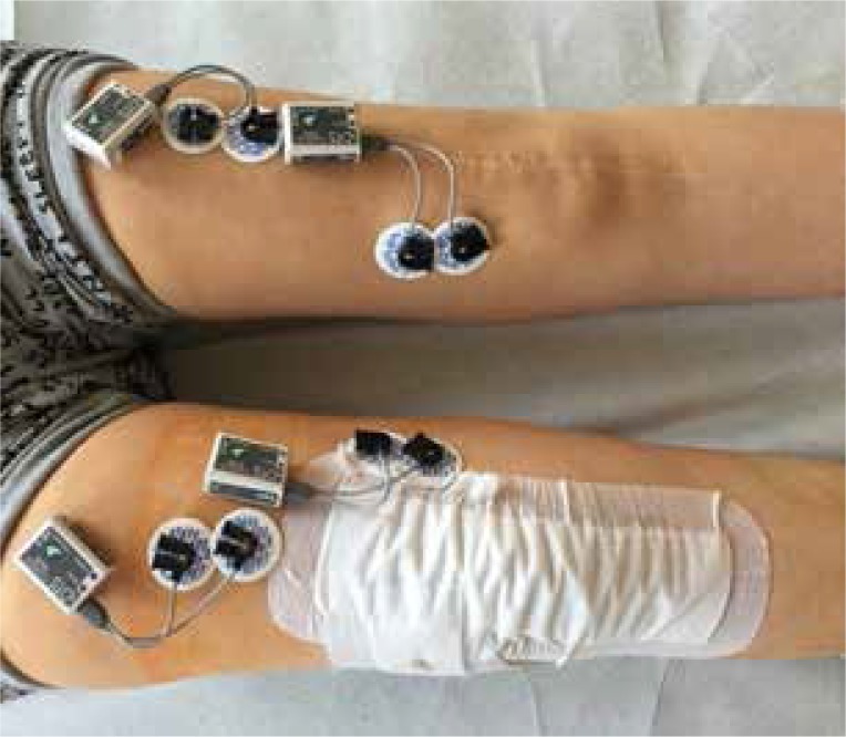
Material and methods: Patients with the diagnosis of rheumatoid arthritis or a degenerative joint disease qualified for surgical treatment were examined. Three groups were analysed: 1) a control group, 2) a group of patients qualified for knee arthroplasty (G1), 3) a group of patients with one knee joint endoprosthesis qualified for the second surgery (G2). The study was carried out through a portable surface electromyography system from Noraxon U.S.A. INC., Clinical DTS and using surface electrodes. The surface electromyography (sEMG) examination was conducted twice: before and on the 10th day after the surgery. The study concerned the quadriceps femoris muscle, i.e. its straight and medial head in both lower limbs during isometric tension and active movement.
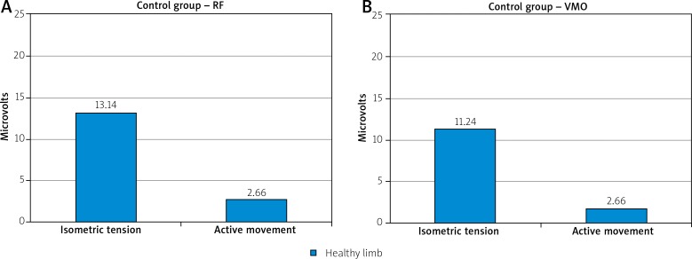
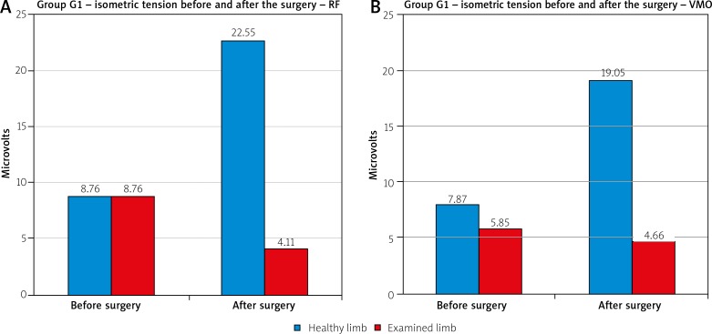
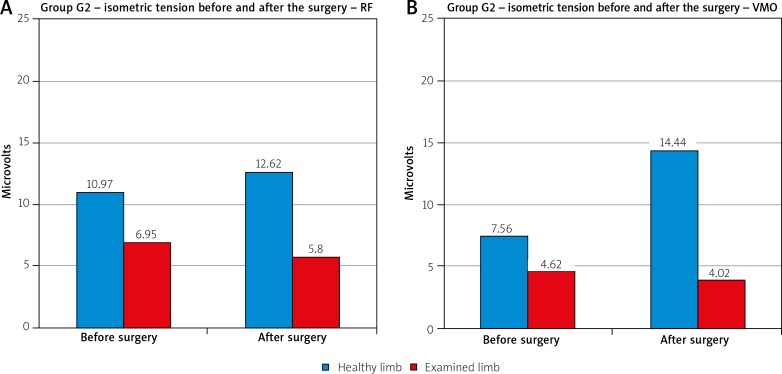
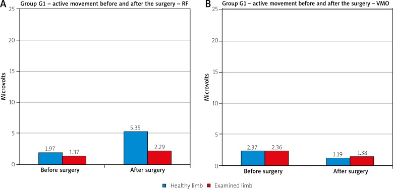
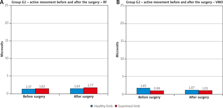
Results: The comparison of the examined muscles' activity in the control group revealed greatly increased activity during isometric tension than during active movement in both muscles. In the G1 group, the comparison of the average values of isometric tension of the examined muscles before the surgery showed slight differences between the healthy limb and the one qualified for the surgical treatment. After the surgery, significant asymmetry between the average values achieved by the healthy and the operated limb could be identified in both muscles. In the G2 group, muscle activity within the currently operated limb revealed only slight differences between the limbs before the surgery. After the surgery, there was an increase in muscle activity within the previously operated limb.
Conclusions: Considerably higher average values of muscle activity during the isometric tension, when compared to the active movement in a sitting position, indicate the necessity of more widespread use of isometric tension in rehabilitating patients after knee arthroplasty.
Keywords: knee joint endoprosthesis; physiotherapy; surface electromyography.
Conflict of interest statement
The authors declare no conflict of interest.
Figures
Similar articles
-
Analysis of Vastus Lateralis and Vastus Medialis Activities in Men the Late Post-Surgery Period after ACL Reconstruction with LARS Synthetic Ligament.Ortop Traumatol Rehabil. 2021 Jun 30;23(3):193-203. doi: 10.5604/01.3001.0014.9157. Ortop Traumatol Rehabil. 2021. PMID: 34187938
-
EMG-angle relationship of the hamstring muscles during maximum knee flexion.J Electromyogr Kinesiol. 2002 Oct;12(5):399-406. doi: 10.1016/s1050-6411(02)00033-0. J Electromyogr Kinesiol. 2002. PMID: 12223173
-
Quadriceps strength asymmetry predicts loading asymmetry during sit-to-stand task in patients with unilateral total knee arthroplasty.Knee Surg Sports Traumatol Arthrosc. 2016 Aug;24(8):2587-94. doi: 10.1007/s00167-015-3827-x. Epub 2015 Oct 8. Knee Surg Sports Traumatol Arthrosc. 2016. PMID: 26450826
-
Contributions to the understanding of gait control.Dan Med J. 2014 Apr;61(4):B4823. Dan Med J. 2014. PMID: 24814597 Review.
-
Rising and sitting down in stroke patients. Auditory feedback and dynamic strength training to enhance symmetrical body weight distribution.Scand J Rehabil Med Suppl. 1994;31:1-57. Scand J Rehabil Med Suppl. 1994. PMID: 7886433 Review.
Cited by
-
The role of electromyography in postoperative total knee arthroplasty: A systematic review.J Orthop. 2025 May 10;65:216-226. doi: 10.1016/j.jor.2025.05.007. eCollection 2025 Jul. J Orthop. 2025. PMID: 40487332
References
-
- Konrad P. ABC of EMG – A Practical Introduction to Kinesiological Electromyography. USA: Noraxon Inc; Version 1.0 April 2005.
-
- Piotrowska SE, Majchrzycki M. Surface Electromyography – A Review. Issue Rehabil Orthop Neurophysiol Sport Promot. 2014;6:19–28.
-
- Karthick PA, Ghosh DM, Swaminathan R. Surface Electromyography Based Muscle Fatigue Detection Using High-Resolution Time-Frequency Methods and Machine Learning Algorithms. Computer Methods and Programs in Biomedicine. 2018;154:45–56. - PubMed
-
- Witkowska A. Importance of the Global Electromyography in Neurophysiological Diagnostics, a Concept of Single Motor Units Action Potentials Analysis (doctoral dissertation) Poznan University of Medical Sciences; 2010.
-
- Sobota G. Electromyography and Its Application in Disorders of the Chewing Apparatus. Dysfunctions of the Chewing Organ. Your Dental Review. 2012;6:57–61.
LinkOut - more resources
Full Text Sources