. 2019 Jan 14:2019:6398296.
doi: 10.34133/2019/6398296. eCollection 2019.

Inside-Out 3D Reversible Ion-Triggered Shape-Morphing Hydrogels

Affiliations

Inside-Out 3D Reversible Ion-Triggered Shape-Morphing Hydrogels

X Du et al. Research (Wash D C). .

Abstract

Shape morphing is a critical aptitude for the survival of organisms and is determined by anisotropic tissue composition and directional orientation of micro- and nanostructures within cell walls, resulting in different swelling behaviors. Recent efforts have been dedicated to mimicking the behaviors that nature has perfected over billions of years. We present a robust strategy for preparing 3D periodically patterned single-component sodium alginate hydrogel sheets cross-linked with Ca2+ ions, which can reversibly deform and be retained into various desirable inside-out shapes as triggered by biocompatible ions (Na+/Ca2+). By changing the orientations of the patterned microchannels or triggering with Na+/Ca2+ ions, various 3D twisting, tubular, and plant-inspired architectures can be facilely programmed. Not only can the transformation recover their initial shapes reversibly, but also it can keep the designated shapes without continuous stimuli. These inside-out 3D reversible ion-triggered hydrogel transformations shall inspire more attractive applications in tissue engineering, biomedical devices, and soft robotics fields.


Conflict of interest statement

The authors declare that they have no conflicts of interest.

Figures

Figure 1
Figure 1
Fabrication and shape deformation of patterned hydrogels. (a) A patterned silica wafer with microchannel structures (width: 800 μm; spacing: 800 μm; height: 100 μm) was fabricated by photolithography. (b) A SA pregel solution was casted onto the silica wafer and dried at room temperature (c). (d) The patterned hydrogel on the silica wafer was first immersed into a 0.1 M CaCl2 solution for 10 min, then cut with microchannels in different orientations after peeling off from the patterned silica wafer (e), and finally immersed into a 0.1 M CaCl2 solution for another 24 h (f). (g) and (h) show the top view and side view of the patterned hydrogel sheet. (i)-(n) show the 3D deformation of the resulting hydrogel sheets with tube-curling structures, helical structures, and rolling structures in the 0.1 M CaCl2 solution and water for 24 h, respectively, with alignment of microchannels at angles θ = 0° (i, l), 45° (j, m), and 90° (k, n) correspondingly. The scale bars are 0.5 cm.
Figure 2
Figure 2
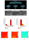
Schematic illustrations and mechanical analyses of the programmed 3D deformations. (a) The scheme shows the differences of the Ca2+ ions diffusion and cross-linking density at different areas of the hydrogel sheet. (b) The cross-sectional field-emission-scanning electron microscopy (FE-SEM) images of the frozen dried hydrogel and the magnified images of the top surface and the bottom surface. (c) Increasing the pre-cross-linking time increases Young's moduli at the three representative areas of the hydrogel sheet. (d) Young's modulus decreases across the thickness from the top surface of the hydrogel sheet, which is 57.55±0.91 MPa for the top surface, 14.94±0.73 MPa for the valley, and 1.42±0.038 MPa for the ridge, respectively. (e) Young's modulus mechanical maps for the top surface, valley (f), and ridge (g), respectively.
Figure 3
Figure 3
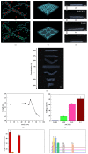
Tunable actuation of programmed hydrogels. (a) In the SA hydrogel, the G-blocks and MG-blocks on polymer chains form ionic cross-links through Ca2+ (b), resulting in tight polymer chains in the CaCl2 solution (c). (e) Partial ionic cross-links are unlocked as certain amounts of Ca2+ ions are replaced by a large amount of Na+ ions, resulting in looser polymer chains in the NaCl solution (f). (d) shows the helical structure of the hydrogel sheet with microchannels facing inward in the 0.1 M CaCl2 solution, which changes its helical rotation oppositely with microchannels facing outward in the 0.1 M NaCl solution (g). (h) The secondly deformed helix can retain its shape in water or recovers to its primary helix with microchannels facing inward again after being immersed in the 0.1 M CaCl2 solution (i). (j) The images of dynamic deformation of hydrogel sheets in a mixed solution of 0.001 M NaCl and 0.001 M CaCl2 for 24 h and the corresponding curve (k). (l) Increasing the concentration of NaCl changes the length of the hydrogel sheet due to the swelling ratio changes in various NaCl solutions (0.0001 M, 0.001 M, 0.01 M, and 0.1 M). (m, n) The EDS demonstrates the change of Na+ and Ca2+ ions in various conditions. The scale bars are 0.5 cm.
Figure 4
Figure 4
Cooperative shape deformations. (a) Connected “T” tube in the 0.1 M CaCl2 solution with combined alignment of microchannels at angles θ = 0° and θ = 90°. (b) Connected “H” tube in the 0.1 M CaCl2 solution with a combined alignment of microchannels at angles θ = 0° and θ = 90°. (c) Connected triangular tube in the 0.1 M CaCl2 solution with combined alignment of microchannels at angles θ = 0°, θ = 60°, and θ = 120°. (d) A double helix in the 0.1 M CaCl2 solution with alignment of microchannels at angle θ = 45°. (e) Connected torsional helix structure in the 0.1 M CaCl2 solution with a combined alignment of microchannels at angles θ = 45° and θ = 135°. (f) Various artificial six-petal flower structures in the 0.1 M CaCl2 solution comprised of microchannels aligned at 0°, 90° (g) and 0°/90° (h). (i) shows the dynamic opening and closing processes of an artificial flower in the 0.1 M NaCl solution, where the petals were partially coated with a blue dye to demonstrate the dynamic inside-out shape transformations of the hydrogels. The scale bars are 1 cm.

References

    1. Ionov L. Biomimetic hydrogel-based actuating systems. Advanced Functional Materials. 2013;23(36):4555–4570. doi: 10.1002/adfm.201203692. - DOI
    1. Poppinga S., Zollfrank C., Prucker O., et al. Toward a new generation of smart biomimetic actuators for architecture. Advanced Materials. 2018;30(19):p. 1703653. doi: 10.1002/adma.201703653. - DOI - PubMed
    1. Wang Z. J., Zhu C. N., Hong W., Wu Z. L., Zheng Q. Cooperative deformations of periodically patterned hydrogels. Science Advances. 2017;3(9):p. e1700348. doi: 10.1126/sciadv.1700348. - DOI - PMC - PubMed
    1. Wang Z. J., Hong W., Wu Z. L., Zheng Q. Site-specific pre-swelling-directed morphing structures of patterned hydrogels. Angewandte Chemie International Edition. 2017;56(50):15974–15978. doi: 10.1002/anie.201708926. - DOI - PubMed
    1. Peng X., Liu T., Zhang Q., Shang C., Bai Q., Wang H. Surface patterning of hydrogels for programmable and complex shape deformations by ion inkjet printing. Advanced Functional Materials. 2017;27(33):p. 1701962. doi: 10.1002/adfm.201701962. - DOI

LinkOut - more resources