

Generating Giant Membrane Vesicles from Live Cells with Preserved Cellular Properties
- PMID: 31549076
- PMCID: PMC6750080
- DOI: 10.34133/2019/6523970
Generating Giant Membrane Vesicles from Live Cells with Preserved Cellular Properties
Abstract
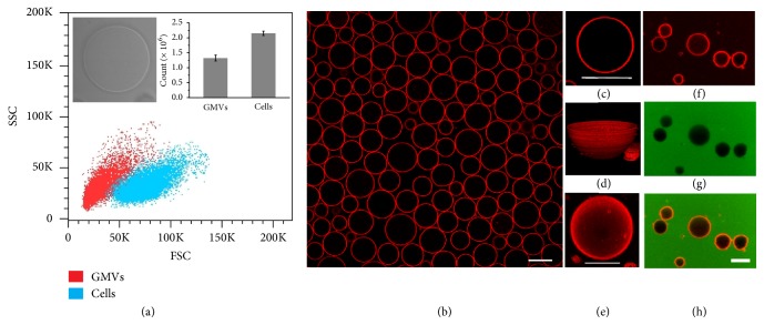
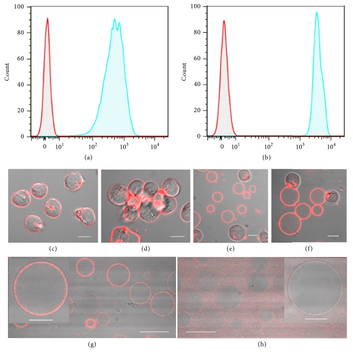
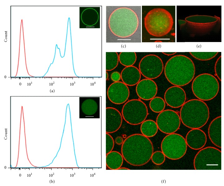
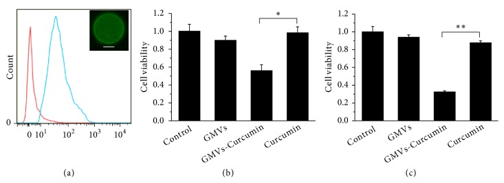
Biomimetic giant membrane vesicles, with size and lipid compositions comparable to cells, have been recognized as an attractive experimental alternative to living systems. Due to the similarity of their membrane structure to that of body cells, cell-derived giant plasma membrane vesicles have been used as a membrane model for studying lipid/protein behavior of plasma membranes. However, further application of biomimetic giant membrane vesicles has been hampered by the side-effects of chemical vesiculants and the utilization of osmotic buffer. We herein develop a facile strategy to derive giant membrane vesicles (GMVs) from mammalian cells in biofriendly medium with high yields. These GMVs preserve membrane properties and adaptability for surface modification and encapsulation of exogenous molecules, which would facilitate their potential biological applications. Moreover, by loading GMVs with therapeutic drugs, GMVs could be employed for drug transport to tumor cells, which represents another step forward in the biomedical application of giant membrane vesicles. This study highlights biocompatible GMVs with biomimicking membrane surface properties and adaptability as an ideal platform for drug delivery strategies with potential clinical applications.
Conflict of interest statement
The authors declare that there are no conflicts of interest regarding the publication of this article.
Figures
Similar articles
-
Biomimetic Carriers Based on Giant Membrane Vesicles for Targeted Drug Delivery and Photodynamic/Photothermal Synergistic Therapy.ACS Appl Mater Interfaces. 2019 Nov 27;11(47):43811-43819. doi: 10.1021/acsami.9b11223. Epub 2019 Nov 18. ACS Appl Mater Interfaces. 2019. PMID: 31670932
-
Membrane Derived Vesicles as Biomimetic Carriers for Targeted Drug Delivery System.Curr Top Med Chem. 2020;20(27):2472-2492. doi: 10.2174/1568026620666200922113054. Curr Top Med Chem. 2020. PMID: 32962615 Review.
-
Proteolipid domains form in biomimetic and cardiac mitochondrial vesicles and are regulated by cardiolipin concentration but not monolyso-cardiolipin.J Biol Chem. 2018 Oct 12;293(41):15933-15946. doi: 10.1074/jbc.RA118.004948. Epub 2018 Aug 29. J Biol Chem. 2018. PMID: 30158245 Free PMC article.
-
Giant multilamellar and large unilamellar lecithin vesicles for the encapsulation and oral delivery of cannabinoids.Food Chem. 2024 Feb 1;433:137291. doi: 10.1016/j.foodchem.2023.137291. Epub 2023 Sep 9. Food Chem. 2024. PMID: 37690133
-
Lipid vesicles in pulsed electric fields: Fundamental principles of the membrane response and its biomedical applications.Adv Colloid Interface Sci. 2017 Nov;249:248-271. doi: 10.1016/j.cis.2017.04.016. Epub 2017 Apr 28. Adv Colloid Interface Sci. 2017. PMID: 28499600 Review.
Cited by
-
Protocells programmed through artificial reaction networks.Chem Sci. 2019 Dec 19;11(3):631-642. doi: 10.1039/c9sc05043d. Chem Sci. 2019. PMID: 34123035 Free PMC article. Review.
-
In Vitro Assays: Friends or Foes of Cell-Penetrating Peptides.Int J Mol Sci. 2020 Jul 2;21(13):4719. doi: 10.3390/ijms21134719. Int J Mol Sci. 2020. PMID: 32630650 Free PMC article. Review.
-
Hydrogel Loaded with Extracellular Vesicles: An Emerging Strategy for Wound Healing.Pharmaceuticals (Basel). 2024 Jul 10;17(7):923. doi: 10.3390/ph17070923. Pharmaceuticals (Basel). 2024. PMID: 39065772 Free PMC article. Review.
-
Cell dehydration enables massive production of engineered membrane vesicles with therapeutic functions.J Extracell Vesicles. 2024 Jul;13(7):e12483. doi: 10.1002/jev2.12483. J Extracell Vesicles. 2024. PMID: 39051765 Free PMC article.
-
Recent progress of artificial cells in structure design, functionality and the prospects in food biotechnology.Mater Today Bio. 2025 Feb 8;31:101565. doi: 10.1016/j.mtbio.2025.101565. eCollection 2025 Apr. Mater Today Bio. 2025. PMID: 40026621 Free PMC article. Review.
References
Grants and funding
LinkOut - more resources
Full Text Sources
