

Rapid Magnetic 3D Printing of Cellular Structures with MCF-7 Cell Inks
- PMID: 31549098
- PMCID: PMC6750075
- DOI: 10.34133/2019/9854593
Rapid Magnetic 3D Printing of Cellular Structures with MCF-7 Cell Inks
Abstract
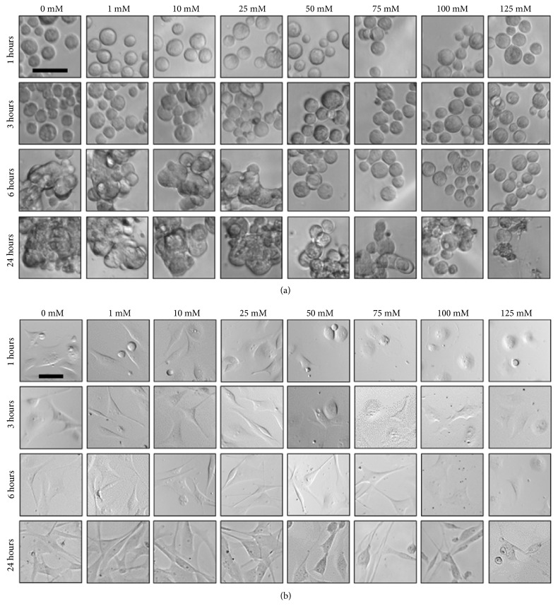
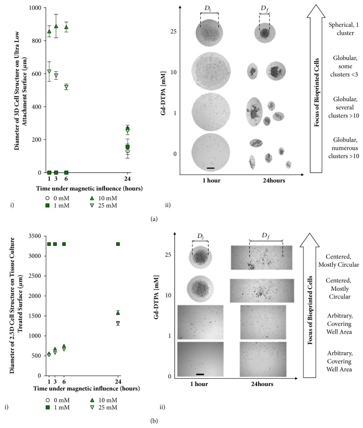
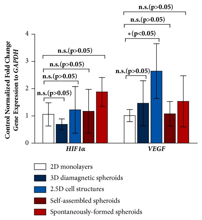
A contactless label-free method using a diamagnetophoretic ink to rapidly print three-dimensional (3D) scaffold-free multicellular structures is described. The inks consist of MCF-7 cells that are suspended in a culture medium to which a paramagnetic salt, diethylenetriaminepentaacetic acid gadolinium (III) dihydrogen salt hydrate (Gd-DTPA), is added. When a magnetic field is applied, the host fluid containing the paramagnetic salt is attracted towards regions of high magnetic field gradient, displacing the ink towards regions with a low gradient. Using this method, 3D structures are printed on ultra-low attachment (ULA) surfaces. On a tissue culture treated (TCT) surface, a 3D printed spheroid coexists with a two-dimensional (2D) cell monolayer, where the composite is termed as a 2.5D structure. The 3D structures can be magnetically printed within 6 hours in a medium containing 25 mM Gd-DTPA. The influence of the paramagnetic salt on MCF-7 cell viability, cell morphology, and ability of cells to adhere to each other to stabilize the printed structures on both ULA and TCT surfaces is investigated. Gene expressions of hypoxia-inducible factor 1-alpha (HIF1α) and vascular endothelial growth factor (VEGF) allow comparison of the relative stresses for the printed 3D and 2.5D cell geometries with those for 3D spheroids formed without magnetic assistance. This magnetic printing method can be potentially scaled to a higher throughput to rapidly print cells into 3D heterogeneous cell structures with variable geometries with repeatable dimensions for applications such as tissue engineering and tumour formation for drug discovery.
Conflict of interest statement
The authors declare that there are no financial conflicts of interest in the regarding the publication of this article.
Figures


Similar articles
-
Fibroblasts Accelerate Formation and Improve Reproducibility of 3D Cellular Structures Printed with Magnetic Assistance.Research (Wash D C). 2020 Jul 23;2020:3970530. doi: 10.34133/2020/3970530. eCollection 2020. Research (Wash D C). 2020. PMID: 32776011 Free PMC article.
-
Suture Fiber Reinforcement of a 3D Printed Gelatin Scaffold for Its Potential Application in Soft Tissue Engineering.Int J Mol Sci. 2021 Oct 27;22(21):11600. doi: 10.3390/ijms222111600. Int J Mol Sci. 2021. PMID: 34769034 Free PMC article.
-
The Effects of Solid Particle Containing Inks on the Printing Quality of Porous Pharmaceutical Structures Fabricated by 3D Semi-Solid Extrusion Printing.Pharm Res. 2022 Jun;39(6):1267-1279. doi: 10.1007/s11095-022-03299-7. Epub 2022 Jun 3. Pharm Res. 2022. PMID: 35661083 Free PMC article.
-
Scaffold-Free Bio-3D Printing Using Spheroids as "Bio-Inks" for Tissue (Re-)Construction and Drug Response Tests.Adv Healthc Mater. 2020 Aug;9(15):e1901831. doi: 10.1002/adhm.201901831. Epub 2020 May 6. Adv Healthc Mater. 2020. PMID: 32378363 Review.
-
Composite Inks for Extrusion Printing of Biological and Biomedical Constructs.ACS Biomater Sci Eng. 2021 Sep 13;7(9):4009-4026. doi: 10.1021/acsbiomaterials.0c01158. Epub 2020 Nov 10. ACS Biomater Sci Eng. 2021. PMID: 34510905 Review.
Cited by
-
3D bioprinted cancer models: from basic biology to drug development.Nat Rev Cancer. 2022 Dec;22(12):679-692. doi: 10.1038/s41568-022-00514-w. Epub 2022 Oct 24. Nat Rev Cancer. 2022. PMID: 36280768 Review.
-
3D-Printed Flat-Bone-Mimetic Bioceramic Scaffolds for Cranial Restoration.Research (Wash D C). 2023 Oct 26;6:0255. doi: 10.34133/research.0255. eCollection 2023. Research (Wash D C). 2023. PMID: 37899773 Free PMC article.
-
Advancement of Scaffold-Based 3D Cellular Models in Cancer Tissue Engineering: An Update.Front Oncol. 2021 Oct 25;11:733652. doi: 10.3389/fonc.2021.733652. eCollection 2021. Front Oncol. 2021. PMID: 34760696 Free PMC article. Review.
-
Three-Dimensional Spheroids as In Vitro Preclinical Models for Cancer Research.Pharmaceutics. 2020 Dec 6;12(12):1186. doi: 10.3390/pharmaceutics12121186. Pharmaceutics. 2020. PMID: 33291351 Free PMC article. Review.
-
Fibroblasts Accelerate Formation and Improve Reproducibility of 3D Cellular Structures Printed with Magnetic Assistance.Research (Wash D C). 2020 Jul 23;2020:3970530. doi: 10.34133/2020/3970530. eCollection 2020. Research (Wash D C). 2020. PMID: 32776011 Free PMC article.
References
-
- Sanyal S. Culture and assay systems used for 3D cell culture. Corning. 2014;9:1–18.
LinkOut - more resources
Full Text Sources
Other Literature Sources
