. 2019 Oct 16;30(10):2528-2532.
doi: 10.1021/acs.bioconjchem.9b00524. Epub 2019 Sep 27.

Assemblies of d-Peptides for Targeting Cell Nucleolus

Affiliations

Assemblies of d-Peptides for Targeting Cell Nucleolus

Huaimin Wang et al. Bioconjug Chem. .

Abstract

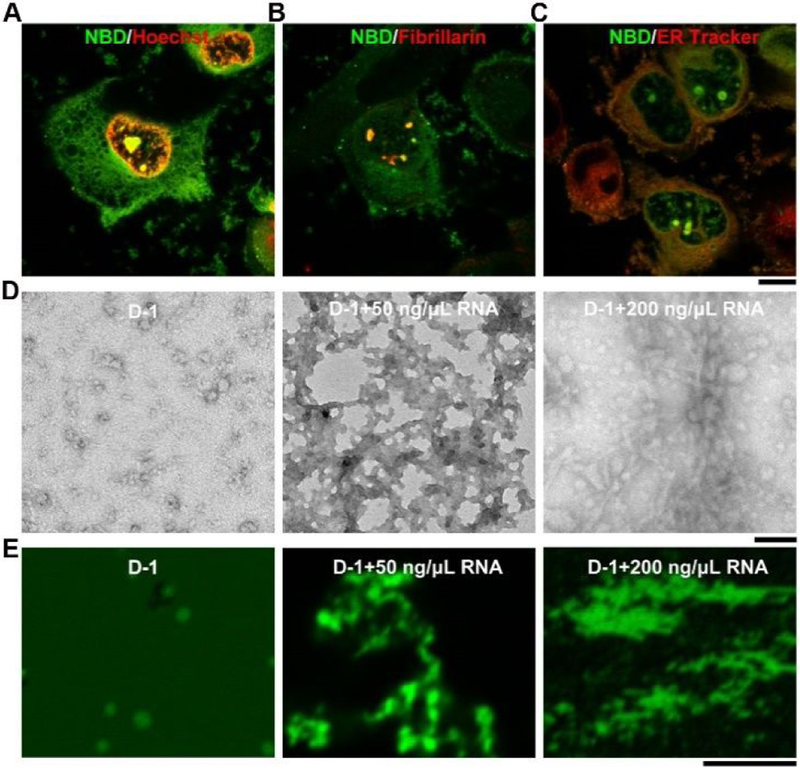
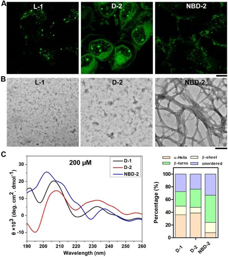
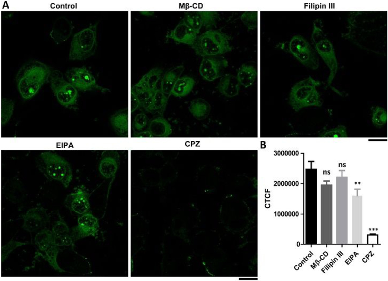
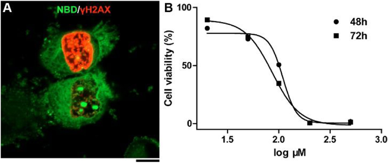
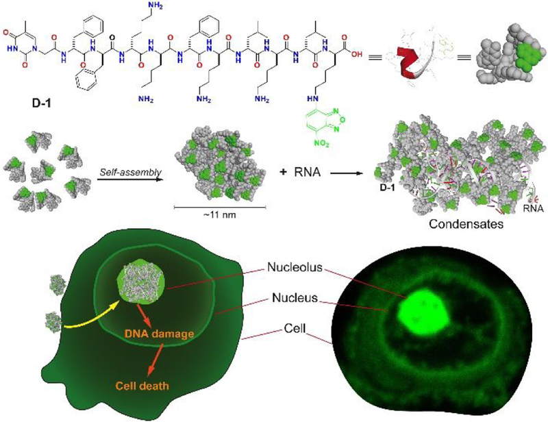
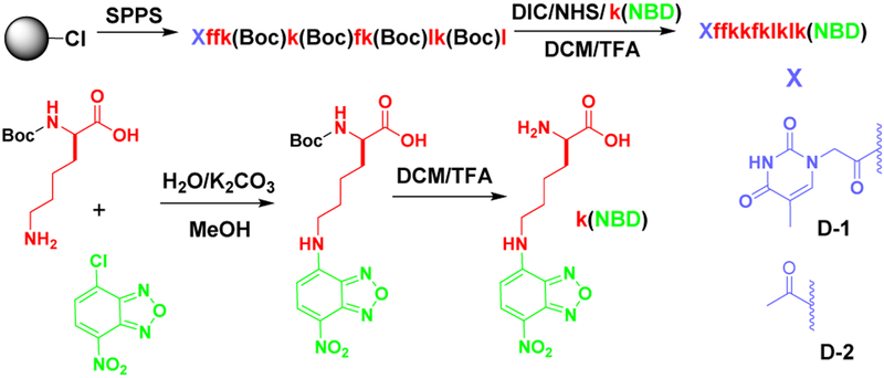
Selectively targeting the cell nucleolus remains a challenge. Here, we report the first case in which d-peptides form membraneless molecular condensates with RNA for targeting cell nucleolus. A d-peptide derivative, enriched with lysine and hydrophobic residues, self-assembles to form nanoparticles, which enter cells through clathrin-dependent endocytosis and mainly accumulate at the cell nucleolus. A structural analogue of the d-peptide reveals that the particle morphology of the assemblies, which depends on the side chain modification, favors the cellular uptake. In contrast to those of the d-peptide, the assemblies of the corresponding l-enantiomer largely localize in cell lysosomes. Preliminary mechanism study suggests that the d-peptide nanoparticles interact with RNA to form membraneless condensates in the nucleolus, which further induces DNA damage and results in cell death. This work illustrates a new strategy for rationally designing supramolecular assemblies of d-peptides for targeting subcellular organelles.


Conflict of interest statement

The authors declare no competing financial interest.

Figures

Figure 1.
Figure 1.
Fluorescent images of T98G cells treated with D-1 for 2 h and then following by (A) Hoechst 33342 staining; (B) immunofluorescence of fibrillarin (abcam, ab5821); (C) ER Tracker staining. Scale bar is 10 μm. (D) TEM and (E) the corresponding confocal laser scanning microscopy (CLSM) images of D-1, and D-1 plus the RNA extracted from T98G cells. All the compounds are dissolved in PBS buffer (final pH=7.4) at concentration of 200 μM. Scale bar in TEM is 100 nm, and 5 μm in CLSM. For better comparing, we changed the color of the Hoechst 33342 to red.
Figure 2.
Figure 2.
Fluorescent images T98G cells treated with (A) L-1, D-2, or NBD-2 for 2 h. Scale bar is 10 μm. (B) TEM images of the assemblies of L-1, D-2, or NBD-2. The concentration of all the molecules is 200 μM. Scale bar is 100 nm. (C) CD spectra of D-1, D-2, and NBD-2 at the concentration of 200 μM, and the percentage of secondary structures at the concentration of 200 μM.
Figure 3.
Figure 3.
(A) Fluorescent images and (B) the corrected total cell fluorescence (CTCF, quantified from the gray scale of CLSM images of 20 cells) of the T98G cells treated with D-1 (200 μM) for 1 h in the absence (control) or presence of the inhibitors M-βCD (5 mM), Filipin III (5 μg/mL), EIPA (100 μM) and CPZ (30 μM). Scale bar is 20 μm. Differences between the group of control and other groups are determined using one-way ANOVA analysis. **: p < 0.005, ***: p < 0.001.
Figure 4.
Figure 4.
(A) Fluorescent image of T98G cells treated with D-1 (200 μM) for 2 h and then analyzed by immunofluorescence of γH2AX. Scale bar is 10 μm. (B) Cytotoxicity of D-1 against T98G cell lines for 48 and 72 h.
Scheme 1.
Scheme 1.
Molecular structures of D-1 and the illustration of membraneless condensates formation of D-1 and RNA.
Scheme 2.
Scheme 2.
Synthetic procedure of D-1 or D-2. Red color and lower case letter in molecular structure represents the D-amino acid residue.

Similar articles

Cited by

References

    1. Shaw P; Jordan E The nucleolus Annu. Rev. Cell Dev. Biol 1995, 11, 93. - PubMed
    1. O’Sullivan JM; Pai DA; Cridge AG; Engelke DR; Ganley AR The nucleolus: a raft adrift in the nuclear sea or the keystone in nuclear structure? Biomol. Concepts 2013, 4, 277. - PMC - PubMed
    1. Raška I; Shaw PJ; Cmarko D Structure and function of the nucleolus in the spotlight Curr. Opin. Cell Biol 2006, 18, 325. - PubMed
    1. Quin JE; Devlin JR; Cameron D; Hannan KM; Pearson RB; Hannan RD Targeting the nucleolus for cancer intervention Biochim. Biophys. Acta, Mol. Basis Dis 2014, 1842, 802. - PubMed
    1. Hein N; Hannan KM; George AJ; Sanij E; Hannan RD The nucleolus: an emerging target for cancer therapy Trends Mol. Med 2013, 19, 643. - PubMed

Publication types