Review
. 2019 Oct;71(4):571-595.
doi: 10.1124/pr.119.017863.

Harnessing Ion-Binding Sites for GPCR Pharmacology

Affiliations
Review

Harnessing Ion-Binding Sites for GPCR Pharmacology

Barbara Zarzycka et al. Pharmacol Rev. 2019 Oct.

Abstract

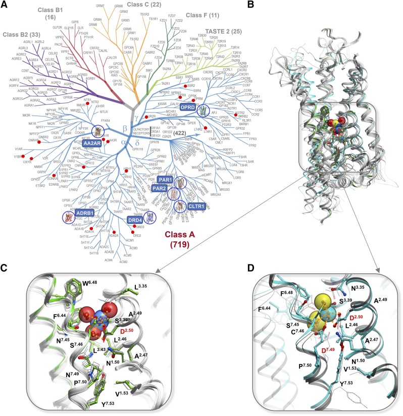
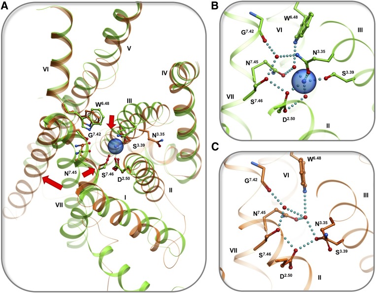
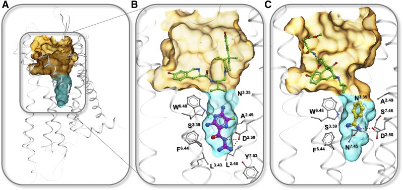
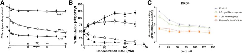
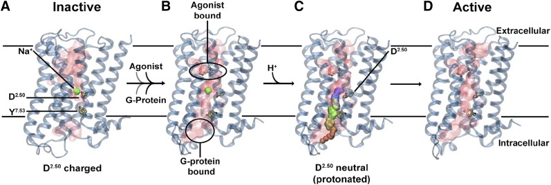
Endogenous ions play important roles in the function and pharmacology of G-protein coupled receptors (GPCRs). Historically the evidence for ionic modulation of GPCR function dates to 1973 with studies of opioid receptors, where it was demonstrated that physiologic concentrations of sodium allosterically attenuated agonist binding. This Na+-selective effect was distinct from effects of other monovalent and divalent cations, with the latter usually counteracting sodium's negative allosteric modulation of binding. Since then, numerous studies documenting the effects of mono- and divalent ions on GPCR function have been published. While ions can act selectively and nonselectively at many sites in different receptors, the discovery of the conserved sodium ion site in class A GPCR structures in 2012 revealed the unique nature of Na+ site, which has emerged as a near-universal site for allosteric modulation of class A GPCR structure and function. In this review, we synthesize and highlight recent advances in the functional, biophysical, and structural characterization of ions bound to GPCRs. Taken together, these findings provide a molecular understanding of the unique roles of Na+ and other ions as GPCR allosteric modulators. We will also discuss how this knowledge can be applied to the redesign of receptors and ligand probes for desired functional and pharmacological profiles. SIGNIFICANCE STATEMENT: The function and pharmacology of GPCRs strongly depend on the presence of mono and divalent ions in experimental assays and in living organisms. Recent insights into the molecular mechanism of this ion-dependent allosterism from structural, biophysical, biochemical, and computational studies provide quantitative understandings of the pharmacological effects of drugs in vitro and in vivo and open new avenues for the rational design of chemical probes and drug candidates with improved properties.


Figures

Fig. 1.
Fig. 1.
Ions identified in GPCR crystal structures. (A) Monovalent ions: Na+ (blue) and Cl (green). (B) Polyvalent ions Zn2+ (magenta), Hg2+ (cyan), PO43− (phosphate colored green), SO42− (sulfur colored yellow). GPCR structures shown as gray cartoons.
Fig. 2.
Fig. 2.
Conserved Na+ sites in GPCRs. (A) GPCR superfamily tree with blue circles highlighting receptor structures with Na+ resolved in the conserved pocket and red dots marking GPCR with established allosteric effect of Na+. (B) Overview of the Na+ position in 7TM helical bundle. (C) Type I Na+ site, first discovered in A2AR (green cartoon and sticks) and conserved in majority of Class A GPCRs structures (gray). (D) Distinct coordination of Na+ by two acidic residues D2.50 and D7.49 in Type II sodium site as resolved in PAR1 (cyan), PAR2 and CLTR1 (gray) in δ-branch GPCRs.
Fig. 3.
Fig. 3.
Conformational changes in the sodium pocket upon activation of opioid receptors. (A) Superimposition of high-resolution structures of δ-opioid receptor in inactive state (green, PDB: 4N6H, resolution 1.8 Å) and μ-opioid receptor in active state (orange, PDB: 5C1M, resolution 2.1 Å); conformational changes shown by arrows. (B) Close up of sodium pocket in inactive state. (C) Close up of the pocket in active state. Hydrogen bonds and salt bridges are shown by cyan dotted lines.
Fig. 4.
Fig. 4.
GPCR pockets for binding allosteric ligands that target conserved sodium binding pocket. Semitransparent surface shows orthosteric pocket (orange) and allosteric conserved sodium pocket (cyan). (A) Overview of the pockets in 7TMD. (B) Amiloride (magenta) bound to KOR in complex with selective antagonist 4-phenylpiperidine derivative JDTic (green) (PDB: 4DJH). (C) Benzamidine (yellow) bound to MOR in complex with irreversible agonist β-FNA (green) (PDB: 4DKL).
Fig. 5.
Fig. 5.
Direct measurement of Na+ effect on GPCR signaling. (A) Exchange of K+ to Na+ leads to reduced basal activity, but maintains agonist-induced signal (Costa et al., 1990). (B) Addition of Na+ dramatically enhances differential between agonists and antagonists of G-protein signaling in MOR (Selley et al., 2000). (C) Addition of Na+ reduces basal activity of DRD4 (Wang et al., 2017). This figure reproduced with permission.
Fig. 6.
Fig. 6.
Updated version of the Na+ involvement in GPR activation mechanism. (A) Inactive receptor conformation has an Na+ ion bound to D2.50 in a pocket, which is sealed from the cytosol by a hydrophobic layer around Y7.53. (B) G-protein and agonist bind to the receptor, leading to the formation of a continuous water channel across the GPCR. The increased mobility of the Na+ ion results in a pKa shift and subsequent protonation of D2.50. (C) Neutralization of D2.50 and the presence of the hydrated pathway facilitate transfer of Na+ to the intracellular side, driven by the TM Na+ gradient and the negative cytoplasmic membrane voltage. (D) The expulsion of Na+ toward the cytosol results in a prolonged active state of the receptor. This figure from Vickery et al. (2018) reproduced with permission.
Fig. 7.
Fig. 7.
GPCR pockets for binding bitopic ligands that target specific ion binding sites. (A) Overview of the pocket positions in 7TM domain. (B) structure of BTL1 receptor with bitopic ligand, PDB: 5X33 (Hori et al., 2018). (C) Model of bitopic ligands designed for binding to KOR Na+ allosteric site (Zaidi et al., 2019). (D) Structure of complex with orthosteric ligand and PO43− ion, PDB: 3RZE (Shimamura et al., 2011). In all panels, semitransparent surface shows orthosteric pocket (orange), allosteric conserved sodium pocket (cyan), and allosteric site in EC loops (green).

Similar articles

Cited by

References

    1. Barchad-Avitzur O, Priest Michael F, Dekel N, Bezanilla F, Parnas H, Ben-Chaim Y. (2016) A novel voltage sensor in the orthosteric binding site of the M2 muscarinic receptor. Biophys J 111:1396–1408. - PMC - PubMed
    1. Batyuk A, Galli L, Ishchenko A, Han GW, Gati C, Popov PA, Lee MY, Stauch B, White TA, Barty A, et al. (2016) Native phasing of x-ray free-electron laser data for a G protein-coupled receptor. Sci Adv 2:e1600292. - PMC - PubMed
    1. Ben-Chaim Y, Chanda B, Dascal N, Bezanilla F, Parnas I, Parnas H. (2006) Movement of ‘gating charge’ is coupled to ligand binding in a G-protein-coupled receptor. Nature 444:106–109. - PubMed
    1. Birdsall NJ, Burgen AS, Hulme EC, Wells JW. (1979) The effects of ions on the binding of agonists and antagonists to muscarinic receptors. Br J Pharmacol 67:371–377. - PMC - PubMed
    1. Birk A, Rinne A, Bunemann M. (2015) Membrane potential controls the efficacy of catecholamine-induced beta1-adrenoceptor activity. J Biol Chem 290:27311–27320. - PMC - PubMed

Publication types

MeSH terms

LinkOut - more resources