Review
. 2019 Nov 1;294(44):15914-15931.
doi: 10.1074/jbc.REV119.009178. Epub 2019 Sep 24.

Solution NMR: A powerful tool for structural and functional studies of membrane proteins in reconstituted environments

Affiliations
Review

Solution NMR: A powerful tool for structural and functional studies of membrane proteins in reconstituted environments

Robbins Puthenveetil et al. J Biol Chem. .

Abstract

A third of the genes in prokaryotic and eukaryotic genomes encode membrane proteins that are either essential for signal transduction and solute transport or function as scaffold structures. Unlike many of their soluble counterparts, the overall structural and functional organization of membrane proteins is sparingly understood. Recent advances in X-ray crystallography, cryo-EM, and nuclear magnetic resonance (NMR) are closing this gap by enabling an in-depth view of these ever-elusive proteins at atomic resolution. Despite substantial technological advancements, however, the overall proportion of membrane protein entries in the Protein Data Bank (PDB) remains <4%. This paucity is mainly attributed to difficulties associated with their expression and purification, propensity to form large multisubunit complexes, and challenges pertinent to identification of an ideal detergent, lipid, or detergent/lipid mixture that closely mimic their native environment. NMR is a powerful technique to obtain atomic-resolution and dynamic details of a protein in solution. This is accomplished through an assortment of isotopic labeling schemes designed to acquire multiple spectra that facilitate deduction of the final protein structure. In this review, we discuss current approaches and technological developments in the determination of membrane protein structures by solution NMR and highlight recent structural and mechanistic insights gained with this technique. We also discuss strategies for overcoming size limitations in NMR applications, and we explore a plethora of membrane mimetics available for the structural and mechanistic understanding of these essential cellular proteins.

Keywords: G-protein–coupled receptor (GPCR); amphipol; bicelle; membrane mimetic; membrane protein; micelle; nanodisc; nanotechnology; nuclear magnetic resonance (NMR); structural biology.


Conflict of interest statement

The authors declare that they have no conflicts of interest with the contents of this article.

Figures

Figure 1.
Figure 1.
Classification of MPs. IMPs with a single TM helix (1); multipass TM helices (2); and β-barrel porin that span both leaflets (3); monotopic amphipathic helix that span a single leaflet (4); lipoproteins (5 and 6); and peripheral extrinsic proteins (7).
Figure 2.
Figure 2.
Micelles. A, spontaneous formation of a micelle from detergents or a mixed micelle with lipids/cholesterol. Also shown are IMPs (α-helical or β-barrel proteins) incorporated in micelles. B, list of popular detergents used for IMPs in solution NMR.
Figure 3.
Figure 3.
Bicelles. A, diameters of spontaneously formed bicelles depend on q values, with higher numbers correlated with wider discs. B, most commonly used lipid, DMPC, which forms the bilayer and detergents, DHPC and CHAPSO, that line the edges. C, spontaneously-oriented bicelles with their bilayer normally perpendicular to the direction of the applied magnetic field; this orientation can be flipped parallel by doping bicelles with paramagnetic ions, including lanthanides.
Figure 4.
Figure 4.
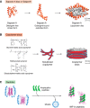
Nanodiscs. A, visual representation of a nanodisc that is either empty or contains IMP. The outer belt protein MSP (green) and lipid molecules are colored by atom type with carbon (gray), oxygen (red), phosphorus (orange), and nitrogen (blue). Also, the approximate hydrocarbon thickness of nanodisc bilayers is shown for various phosphatidylcholines. B, schematic representation of the overall length and disc diameters for several MSP variants. Nanodiscs with smaller diameters (below 8.5 nm) are preferable for solution NMR studies. The largest “MACRODISC,” obtained from a 14-amino acid peptide, produces a disc of 30 nm diameter and serves as an alignment medium.
Figure 5.
Figure 5.
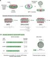
Other potentially useful membrane mimetics. A, saposin-A in its detergent-free form adopts a closed conformation that becomes extended when bound to detergent molecules (PDB code 4DDJ). Saposin-A lipoprotein disc, with a diameter of 3.2 nm, contains two saposin-A proteins brought together by a lipid core. B, SMALP is shown where the synthetic styrene–maleic acid co-polymer forms discs by encapsulating lipid within its central cavity. SMA lipid discs or Lipodisq® has a diameter of 10 nm. Other polymers shown to form lipid particles include methacrylate and DIBMA. C, amphipathic peptides have been recently used for “lipid-free” IMP reconstitution forming “peptidisc”: helical peptides wrap around the hydrophobic parts of detergent-purified IMPs eventually displacing detergent molecules.
Figure 6.
Figure 6.
Comprehensive overview of solution NMR IMP structures in PDB as of June, 2019.
Figure 7.
Figure 7.
Applicability of nanodisc systems for studying signaling pathways. The cytoplasmic tail of integrin β3 subunit is phosphorylated by Src kinase (in vitro) in an NMR tube: 15N-labeled β3 incorporated discs were mixed with the kinase domain of Src kinase in the presence of ATP. Phosphorylation is manifested through chemical shift perturbations observed in the overlay of the 1H–15N TROSY HSQC spectra obtained from the unphosphorylated (black) and bi-phosphorylated (red) β3 were collected on a 600-MHz magnet. Also shown is the β3 tail sequence indicating phosphorylation sites at the two tyrosine residues.
Figure 8.
Figure 8.
Illustration with a few examples of structural discrepancies.

Similar articles

Cited by

References

    1. Drews J. (2000) Drug discovery: a historical perspective. Science 287, 1960–1964 10.1126/science.287.5460.1960 - DOI - PubMed
    1. Lundstrom K. (2009) An overview on GPCRs and drug discovery: structure-based drug design and structural biology on GPCRs. Methods Mol. Biol. 552, 51–66 10.1007/978-1-60327-317-6_4 - DOI - PMC - PubMed
    1. Ernst R. R., Bodenhausen G., and Wokaun A. (1987) Principles of Nuclear Magnetic Resonance in One and Two Dimensions, Oxford University Press, Oxford, UK
    1. Wuthrich K. (1986) NMR of Proteins and Nucleic Acids, John Wiley & Sons, New York
    1. Oschkinat H., Griesinger C., Kraulis P. J., Sørensen O. W., Ernst R. R., Gronenborn A. M., and Clore G. M. (1988) Three-dimensional NMR spectroscopy of a protein in solution. Nature 332, 374–376 10.1038/332374a0 - DOI - PubMed

Associated data

LinkOut - more resources