. 2019 Sep 24;9(1):13803.
doi: 10.1038/s41598-019-50023-4.

Long-term mucosal injury and repair in a murine model of pelvic radiotherapy

Affiliations

Long-term mucosal injury and repair in a murine model of pelvic radiotherapy

Dilip K Malipatlolla et al. Sci Rep. .

Abstract

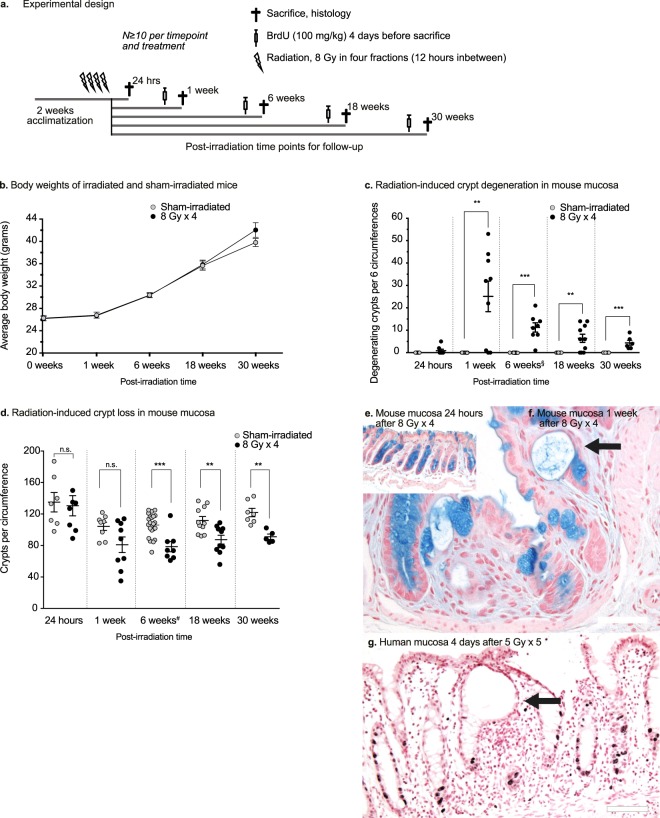
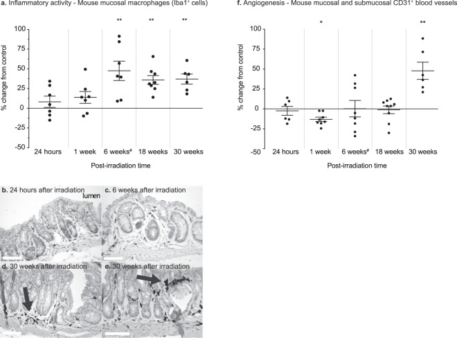
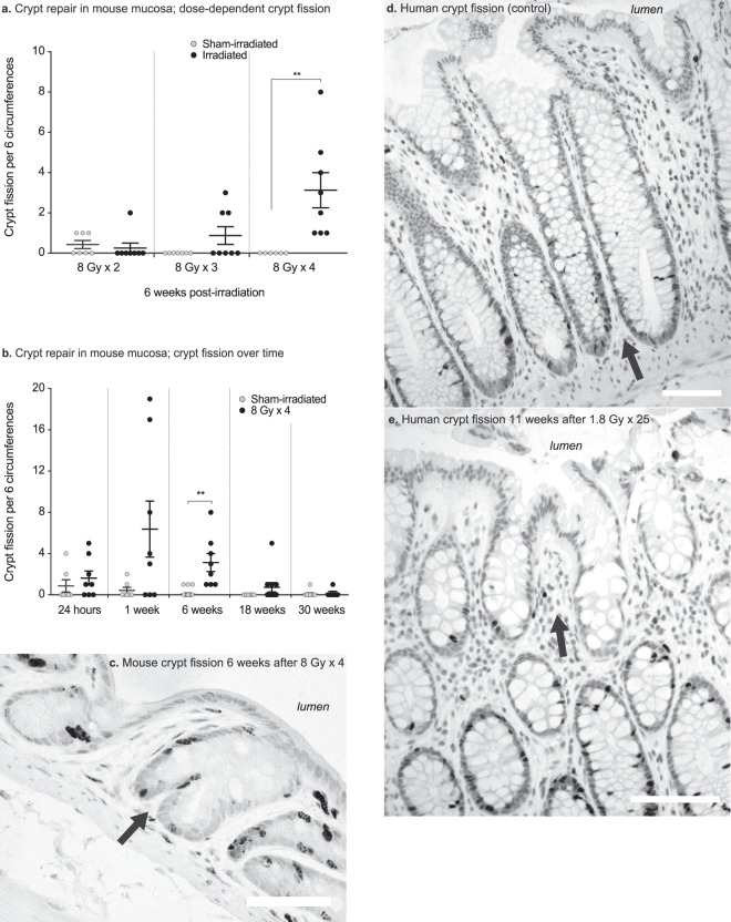
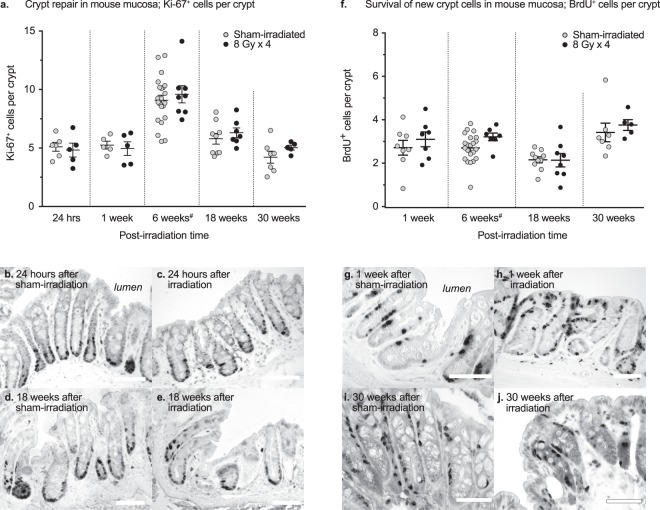
Chronic intestinal injury after pelvic radiotherapy affects countless cancer survivors worldwide. A comprehensive understanding of the long-term injury dynamics is prevented in available animal models. With linear accelerators that are used to treat cancer in patients, we irradiated a small volume encompassing the colorectum in mice with four fractions of 8 Gy per fraction. We then determined the long-term dynamics of mucosal injury, repair, and the duration of inflammation. We show that crypt fission, not cell proliferation, is the main long-term mechanism for rescuing crypt density after irradiation, and provides a potentially wide window for clinical interventions. Persisting macrophage aggregations indicate a chronic mucosal inflammation. A better understanding as to how crypt fission is triggered and why it fails to repair fully the mucosa may help restore bowel health after pelvic radiotherapy. Moreover, anti-inflammatory interventions, even if implemented long after completed radiotherapy, could promote bowel health in pelvic cancer survivors.


Conflict of interest statement

The authors declare no competing interests.

Figures

Figure 1
Figure 1
(a) Experimental design. Male C57BL/6J mice (N ≧ 10 per treatment and time point) were irradiated with four fractions of 8 Gy (8 Gy × 4) with 12 hours between each delivered fraction. Four days before sacrifice, the mice were injected with BrdU (100 mg/kg), to follow crypt cell survival. Colorectal tissues were harvested at 24 hours, 1 week, 6 weeks, 18 weeks, and 30 weeks post-irradiation. (b) Weight curves (grams) for the 30-week group. The mice survived and gained weight in a manner similar to the sham-irradiated controls. At 30 weeks post-irradiation, there is a trend towards increased weight in the irradiated mice. (c) Number of degenerating crypts per six circumferences. Radiation-induced crypt degeneration is evident first at 1 week post-irradiation. The number of degenerating crypts diminishes over time. (d) Crypt loss over time. Quantification of the surviving crypts per circumference of the colorectum reveals that the crypt loss is permanent. At 24 hours post-irradiation, no crypt loss is detected. One week after irradiation, a few animals have lost more than 50% of their crypts per circumference, as compared to the sham-irradiated controls, while some animals still have the normal number of crypts. At 6 weeks post-irradiation, crypt loss is seen in nearly all the animals in the irradiated group. The irradiated mucosa has approximately 25% fewer crypts than the sham-irradiated mucosa, and this remains unchanged throughout the experiment, despite what appears to be a slight increase in the number of crypts with age in both groups. §,#Data from the 6-week group have been reported previously, and, respectively. (e,f) Histology of irradiated mucosa samples. There are negligible numbers of degenerating crypts 24 hours after irradiation (e). However, at 1 week post-irradiation, several animals have multiple degenerating crypts (f, arrow) with few goblet cells (Alcian Blue and Nuclear Fast Red stain). Scale bars, 50 μm. (g) Degenerating crypt in the rectal mucosa of a cancer patient 4 days after 5 Gy × 5 (G, arrow). There are multiple dividing cells in nearby crypts, visualised as darkly stained Ki-67+ nuclei. Only two Ki-67+ cells are visible at the bottom of the degenerating crypt. Scale bar, 100 μm.
Figure 2
Figure 2
(a) Percentage change (relative to sham-irradiated animals) in the number of Iba1+ mucosal macrophages at 24 hours, 1 week, 6 weeks, 18 weeks, and 30 weeks post-irradiation. An increase in infiltrating macrophages is not seen until 6 weeks after irradiation, with most of the animals exhibiting an increase in the number of mucosal macrophages. This remains unchanged over time, indicating a state of long-lasting, possibly chronic mucosal inflammation. (be) Iba1+-expressing macrophages visualised with DAB immunohistochemistry in the irradiated colorectal mucosa at various time-points after irradiation. Macrophages are still abundant in the mucosa 30 weeks after irradiation and tend to aggregate around the blood vessels, close to the degenerating crypts, and under the epithelial surface (arrows). Scale bars, 50 μm. (f) CD31+ blood vessels in irradiated animals, shown in terms of the percentage change relative to the sham-irradiated animals. The increase in blood vessels at 30 weeks after irradiation suggests late, compensatory angiogenesis. #The data for the 6-week group have been published previously.
Figure 3
Figure 3
(a) Numbers of crypt fission events per 6 circumferences after different fractionation schedules. The occurrence of crypt fission is dose-dependent, as seen 6 weeks after irradiation. (b) Number of crypt fission events per six circumferences at 24 hours, and at 1 week, 6 weeks, 18 weeks, and 30 weeks after irradiation or sham-irradiation. A few of the irradiated animals show many crypt fission events 1 week after irradiation, and at 6 weeks post-irradiation all the animals exhibit crypt fission; thereafter the number of fission events declines. At 30 weeks post-irradiation, there is no difference in the number of crypt fissions between the irradiated and sham-irradiated animals. (c) Irradiated mouse colorectal mucosa 6 weeks after 8 Gy × 4 of irradiation. The dark nuclei are cells that are labelled with BrdU, showing newly born cells migrating to form two new crypt walls at the crypt mid-line (arrow). (d) Crypt fission in a non-irradiated human rectal biopsy (arrow), showing multiple darkly stained Ki-67+ proliferating cells at the bottom of the dividing crypt. (e) Crypt fission in a human rectal biopsy (arrow) harvested 11 weeks after 1.8 Gy × 25. A few Ki-67+ proliferating cells are seen at the bottom of one of the two resulting crypts.
Figure 4
Figure 4
(a) No statistically significant change in the numbers of Ki-67-labelled cells is observed after irradiation at any of the time-points chosen. (be) Numbers of Ki-67+ cells at 24 hours (b,c) and 18 weeks (d,e) post-irradiation, showing similar numbers of proliferating cells in the surviving crypts of irradiated animals, as compared to sham-irradiated animals. (f) Percentage change (relative to sham-irradiated animals) in the number of surviving cells per crypt, as evidenced by BrdU-labelling. The rate of cell survival remains unchanged at 24 hours after irradiation and thereafter. (gj) BrdU+ crypt cells in irradiated and sham-irradiated mucosal samples after 1 week and 30 weeks. Scale bars, 50 μm. #Data for the 6-week group have been previously published.

References

    1. Andreyev HJ, Wotherspoon A, Denham JW, Hauer-Jensen M. Defining pelvic-radiation disease for the survivorship era. Lancet Oncol. 2010;11:310–312. doi: 10.1016/S1470-2045(10)70026-7. - DOI - PubMed
    1. Dunberger G, et al. Fecal incontinence affecting quality of life and social functioning among long-term gynecological cancer survivors. Int J Gynecol Cancer. 2010;20:449–460. doi: 10.1111/IGC.0b013e3181d373bf. - DOI - PubMed
    1. Steineck G, et al. Identifying radiation-induced survivorship syndromes affecting bowel health in a cohort of gynecological cancer survivors. PLoS One. 2017;12:e0171461. doi: 10.1371/journal.pone.0171461. - DOI - PMC - PubMed
    1. Potten CS, Grant HK. The relationship between ionizing radiation-induced apoptosis and stem cells in the small and large intestine. Br J Cancer. 1998;78:993–1003. doi: 10.1038/bjc.1998.618. - DOI - PMC - PubMed
    1. Mihaescu A, et al. Critical role of P-selectin and lymphocyte function antigen-1 in radiation-induced leukocyte-endothelial cell interactions in the colon. Dis Colon Rectum. 2007;50:2194–2202. doi: 10.1007/s10350-007-9065-7. - DOI - PubMed

Publication types