. 2021 Apr;18(4):1045-1057.
doi: 10.1038/s41423-019-0296-z. Epub 2019 Sep 24.

NLRP3 inflammasome blockade reduces adipose tissue inflammation and extracellular matrix remodeling

Affiliations

NLRP3 inflammasome blockade reduces adipose tissue inflammation and extracellular matrix remodeling

Xabier Unamuno et al. Cell Mol Immunol. 2021 Apr.

Abstract

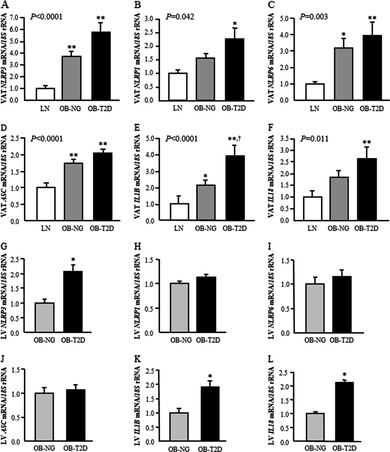
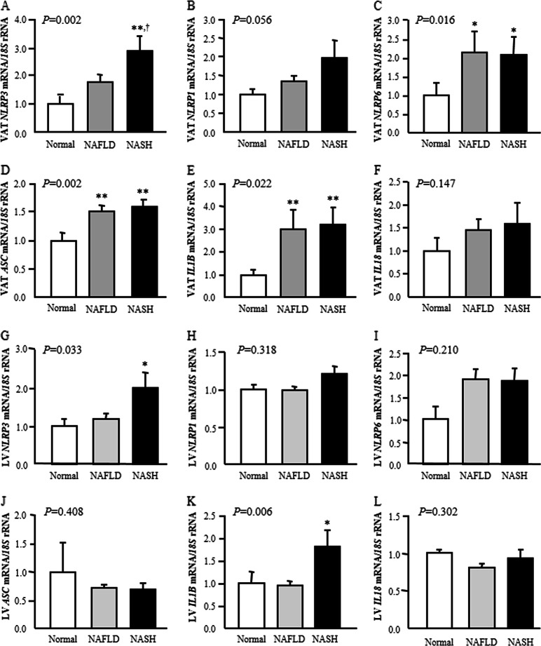
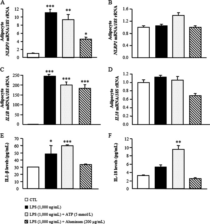
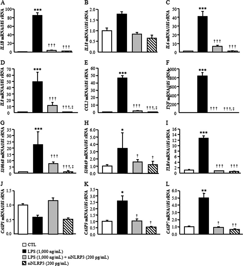
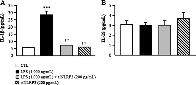
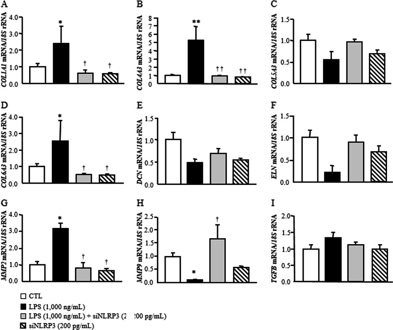
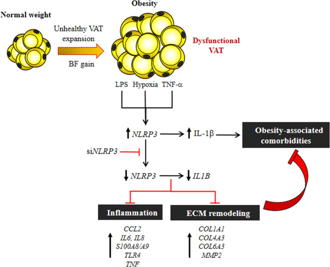
The NLRP3-IL-1β pathway plays an important role in adipose tissue (AT)-induced inflammation and the development of obesity-associated comorbidities. We aimed to determine the impact of NLRP3 on obesity and its associated metabolic alterations as well as its role in adipocyte inflammation and extracellular matrix (ECM) remodeling. Samples obtained from 98 subjects were used in a case-control study. The expression of different components of the inflammasome as well as their main effectors and inflammation- and ECM remodeling-related genes were analyzed. The impact of blocking NLRP3 using siRNA in lipopolysaccharide (LPS)-mediated inflammation and ECM remodeling signaling pathways was evaluated. We demonstrated that obesity (P < 0.01), obesity-associated T2D (P < 0.01) and NAFLD (P < 0.05) increased the expression of different components of the inflammasome as well as the expression and release of IL-1β and IL-18 in AT. We also found that obese patients with T2D exhibited increased (P < 0.05) hepatic gene expression levels of NLRP3, IL1B and IL18. We showed that NLRP3, but not NLRP1, is regulated by inflammation and hypoxia in visceral adipocytes. We revealed that the inhibition of NLRP3 in human visceral adipocytes significantly blocked (P < 0.01) LPS-induced inflammation by downregulating the mRNA levels of CCL2, IL1B, IL6, IL8, S100A8, S100A9, TLR4 and TNF as well as inhibiting (P < 0.01) the secretion of IL1-β into the culture medium. Furthermore, blocking NLRP3 attenuated (P < 0.01) the LPS-induced expression of important molecules involved in AT fibrosis (COL1A1, COL4A3, COL6A3 and MMP2). These novel findings provide evidence that blocking the expression of NLRP3 reduces AT inflammation with significant fibrosis attenuation.

Keywords: Inflammasone; Inflammation; NLRP3; Nonalcoholic fatty liver disease; Obesity; Type 2 diabetes.


Conflict of interest statement

The authors declare no competing interests.

Figures

Fig. 1
Fig. 1
Gene expression levels in visceral adipose tissue and liver of the main inflammasome components in obesity and obesity-associated type 2 diabetes. Bar graphs show the mRNA levels of a, g NLRP3, b, h NLRP1, c, i NLRP6, d, j ASC, e, k IL1B and f, l IL18 in visceral adipose tissue and liver, respectively, from lean volunteers (LN), obese NG subjects (OB-NG) and obese patients with T2D (OB-T2D). Bars represent the mean ± SEM. Differences between groups were analyzed by one-way ANOVA followed by Tukey’s tests. *P < 0.05 and **P < 0.01 vs. lean subjects. P < 0.05 vs. OB-NG
Fig. 2
Fig. 2
Effect of nonalcoholic fatty liver disease (NAFLD) and nonalcoholic steatohepatitis (NASH) on visceral adipose tissue and hepatic gene expression levels of the main inflammasome components. Bar graphs show the mRNA levels of a, g NLRP3, b, h NLRP1, c, i NLRP6, d, j ASC, e, k IL1B and f, l IL18 in visceral adipose tissue and liver according to hepatic function. Bars represent the mean ± SEM. Differences between groups were analyzed by one-way ANOVA followed by Tukey’s tests. *P < 0.05 and **P < 0.01 vs. normal liver. P < 0.05 vs. NAFLD
Fig. 3
Fig. 3
LPS alone or combined with ATP or aluminum promotes the expression and secretion of IL-1β by priming the expression of the NLRP3 inflammasome in human visceral adipocytes. Gene expression levels of a NLRP3, b NLRP1, c IL1B and d IL18 as well as released levels of e IL-1β and f IL-18 in cultured human visceral adipocytes incubated with LPS (1000 ng/mL) for 24 h as well as with LPS (1000 ng/mL) for 3 h in the presence or absence of ATP (5 mmol/L) and aluminum (200 μg/mL) for another 24 h. Gene expression levels in unstimulated cells were assumed to be 1. Values are the mean ± SEM (n = 6 per group). Differences between groups were analyzed by one-way ANOVA followed by Dunnett’s tests. *P < 0.05, **P < 0.01 and ***P < 0.001 vs. unstimulated cells
Fig. 4
Fig. 4
Knockdown of NLRP3 ameliorated LPS-induced inflammation in human visceral adipocytes. Gene expression levels of inflammatory factors in human visceral adipocytes incubated in the presence or absence of LPS (1000 ng/mL) for 3 h, followed by transfection with or without 100 pmol/L NLRP3 siRNA/2 × 105 cells/well for another 24 h. Gene expression levels in scrambled siRNA cells (CTL) were assumed to be 1. Values are the mean ± SEM (n = 6 per group). Differences between groups were analyzed by one-way ANOVA followed by Tukey’s tests. *P < 0.05, **P < 0.01 and ***P < 0.001 vs. scrambled siRNA. P < 0.05, ††P < 0.01 and †††P < 0.001 vs. adipocytes stimulated with LPS. P < 0.05 vs. adipocytes stimulated with LPS and siNLRP3
Fig. 5
Fig. 5
Secreted levels of IL-1β and IL-18 after NLRP3 siRNA treatment in human visceral adipocytes. a IL-1β and b IL-18 concentrations in the culture media of human visceral adipocytes incubated in the presence or absence of LPS (1000 ng/mL) for 3 h, followed by transfection with or without 100 pmol/L NLRP3 siRNA/2 × 105 cells/well for another 24 h. Values are the mean ± SEM (n = 6 per group). Differences between groups were analyzed by one-way ANOVA followed by Tukey’s tests. ***P < 0.001 vs. unstimulated cells. ††P < 0.01 vs. adipocytes stimulated with LPS
Fig. 6
Fig. 6
Effect of blocking NLRP3 on the LPS-induced expression of extracellular matrix remodeling genes in human visceral adipocytes. Gene expression levels of fibrosis-related factors in human visceral adipocytes incubated in the presence or absence of LPS (1000 ng/mL) for 3 h, followed by transfection with or without 100 pmol/L NLRP3 siRNA/2 × 105 cells/well for another 24 h. Gene expression levels in scrambled siRNA cells (CTL) were assumed to be 1. Values are the mean ± SEM (n = 6 per group). Differences between groups were analyzed by one-way ANOVA followed by Tukey’s tests. *P < 0.05 and **P < 0.01 vs. scrambled siRNA. P < 0.05 and ††P < 0.01 vs. adipocytes stimulated with LPS
Fig. 7
Fig. 7
Obesity-associated AT dysfunction favors the development of different comorbidities, including T2D and NAFLD. Danger signals, including endogenous (TNF-α) and exogenous (LPS) inflammatory factors as well as hypoxia, are able to induce the expression of the cytosolic innate immune signaling receptor NLRP3 and subsequently the activation of IL-1β. NLRP3 silencing using siRNA inhibits the expression and release of IL-β as well as important factors promoting inflammation (CCL2, IL6, IL8, S100A8, S100A9 and TNF) and fibrosis (COL1A1, COL4A3, COL6A3 and MMP2) in human visceral adipocytes. In this sense, direct targeting of NLRP3 could offer potential efficacy in the treatment of obesity-associated comorbidities

References

    1. GBDO Collaborators, et al. Health effects of overweight and obesity in 195 countries over 25 years. N. Engl. J. Med. 2017;377:13–27. - PMC - PubMed
    1. James WPT. Obesity: a global public health challenge. Clin. Chem. 2018;64:24–29. - PubMed
    1. Lee YS, Wollam J, Olefsky JM. An integrated view of immunometabolism. Cell. 2018;172:22–40. - PMC - PubMed
    1. Bray GA, Frühbeck G, Ryan DH, Wilding JP. Management of obesity. Lancet. 2016;387:1947–1956. - PubMed
    1. Ouchi N, Parker JL, Lugus JJ, Walsh K. Adipokines in inflammation and metabolic disease. Nat. Rev. Immunol. 2011;11:85–97. - PMC - PubMed

Publication types

MeSH terms