Review
. 2019 Dec;26(12):2495-2512.
doi: 10.1038/s41418-019-0411-9. Epub 2019 Sep 24.

Translating neural stem cells to neurons in the mammalian brain

Affiliations
Review

Translating neural stem cells to neurons in the mammalian brain

Siraj K Zahr et al. Cell Death Differ. 2019 Dec.

Abstract

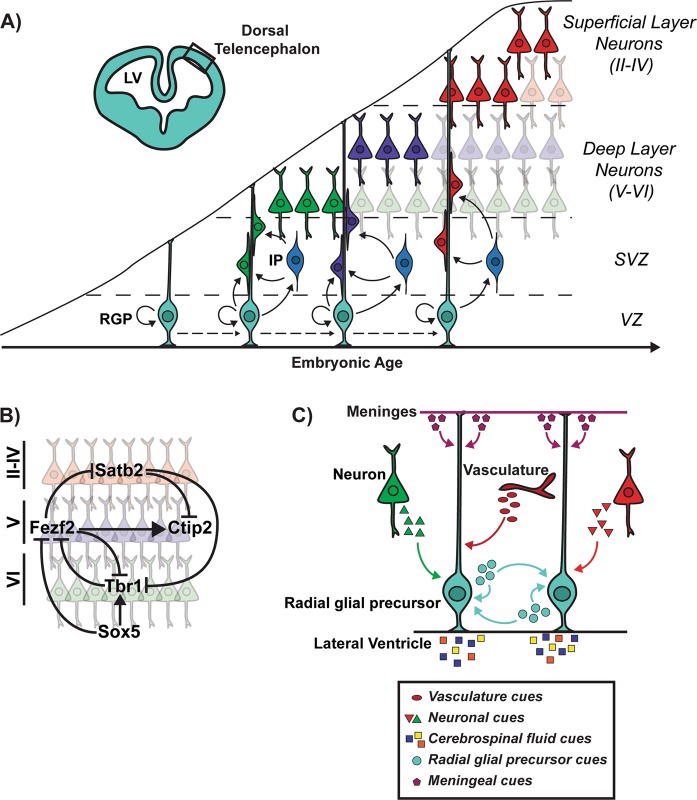
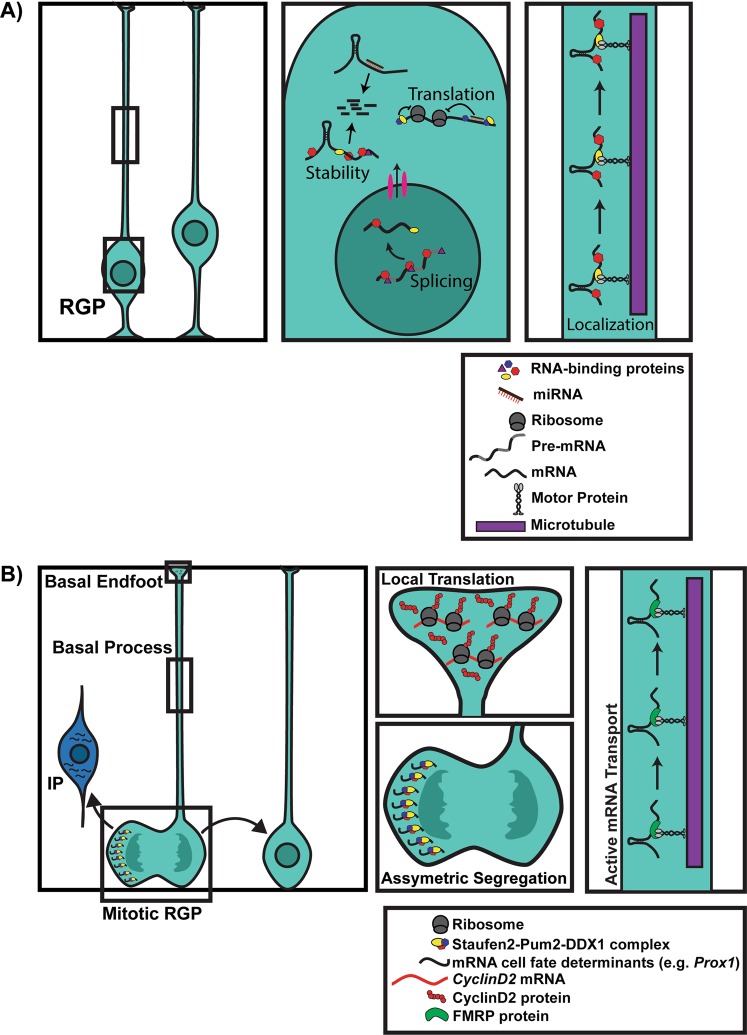
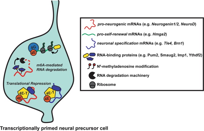
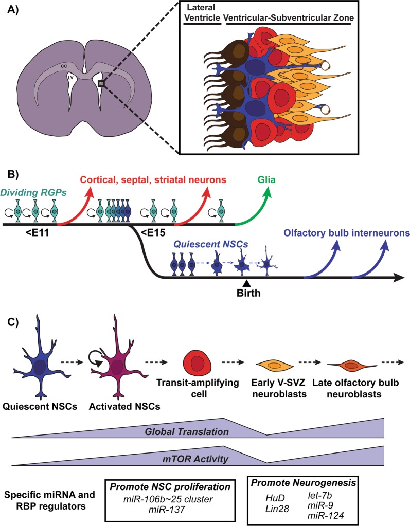
The mammalian neocortex underlies our perception of sensory information, performance of motor activities, and higher-order cognition. During mammalian embryogenesis, radial glial precursor cells sequentially give rise to diverse populations of excitatory cortical neurons, followed by astrocytes and oligodendrocytes. A subpopulation of these embryonic neural precursors persists into adulthood as neural stem cells, which give rise to inhibitory interneurons and glia. Although the intrinsic mechanisms instructing the genesis of these distinct progeny have been well-studied, most work to date has focused on transcriptional, epigenetic, and cell-cycle control. Recent studies, however, have shown that posttranscriptional mechanisms also regulate the cell fate choices of transcriptionally primed neural precursors during cortical development. These mechanisms are mediated primarily by RNA-binding proteins and microRNAs that coordinately regulate mRNA translation, stability, splicing, and localization. Together, these findings point to an extensive network of posttranscriptional control and provide insight into both normal cortical development and disease. They also add another layer of complexity to brain development and raise important biological questions for future investigation.


Conflict of interest statement

The authors declare that they have no conflict of interest.

Figures

Fig. 1
Fig. 1
Embryonic cortical neurogenesis occurs in an inside-out fashion. a In the developing dorsal telencephalon, radial glial precursors (RGPs) residing in the ventricular zone (VZ) adjacent to the lateral ventricles (LV) can give rise to a postmitotic neuron directly or indirectly via an intermediate progenitor (IP) cell. IPs populate the space basal to the VZ known as the subventricular zone (SVZ). Cortical neurons are generated in an inside-out fashion to populate the six layers of the cortex. Earlier-born neurons (shown in green and purple) populate the deepest of the six cortical layers (V–VI), while later born neurons (shown in red) populate progressively more superficial layers (II–IV). These layers contain distinct neuronal subtypes that differ based on morphology, electrophysiological activity, axonal connectivity, and gene expression. b Illustration of a cross-repressive transcriptional circuit that regulates deep versus superficial layer neuron specification [1, 4]. Tbr1 specifies deep layer VI corticothalamic neurons (shown in green) in part by repressing Fezf2, while Fezf2 acts upstream of Ctip2 to specify deep layer V subcerebral neurons (shown in purple) [1, 4]. Sox5 regulates the timing of deep layer neurogenesis by repressing Fezf2 until the production of layer VI corticothalamic neurons is complete. Satb2 specifies upper layer neurons (shown in red) in part by repressing deep-layer neuronal specifiers Ctip2 and Tbr1 [1, 4]. c RGPs integrate autocrine and paracrine factors originating from several sources including the meninges, vasculature, newborn neurons, and cerebrospinal fluid (CSF), many of which regulate RGP cell fate decisions
Fig. 2
Fig. 2
NPC activity is dynamically regulated by posttranscriptional mechanisms. a Radial glial precursors (RGPs, light blue) are dynamically regulated by posttranscriptional mechanisms. The boxed regions of the RGP cell body and basal process are shown at higher magnification at the right. RBPs (shown by colored hexagons, triangles, and ovals) and miRNAs (red/brown lines) are highly ubiquitous and influence various steps of RNA metabolism including mRNA splicing, nuclear export, stability, localization, and translation. In the basal process, mRNAs are actively transported by RBPs (such as FMRP) along microtubules (purple). A figure legend is shown in the lower right panel. b mRNA cell fate determinants are asymmetrically segregated and actively transported in RGPs to ensure appropriate cell fate decisions. In RGPs undergoing mitosis, a Staufen2-Pum2-Ddx1 complex asymmetrically segregates cell fate determinants such as Prox1 mRNA into the daughter cell destined to become an intermediate progenitor (IP). mRNA cell fate determinants are actively transported to basal endfeet by FMRP, where they are locally translated. In RGP basal endfeet, self-renewal factors such as CyclinD2 mRNA are locally translated, ensuring that the daughter cell inheriting the basal process maintains its self-renewing capacity. A figure legend is shown in the lower right panel
Fig. 3
Fig. 3
Transcriptional priming and posttranscriptional control in embryonic neural precursor cells (NPCs). Embryonic NPCs are transcriptionally primed to differentiate into diverse neuronal progeny, but maintained in an undifferentiated state until the appropriate time via posttranscriptional mechanisms. These mechanisms include degradation of m6A-modified mRNAs mediated by RNA-binding proteins (RBPs) such as Ythdf2 and translational repression by a Pum2-4E-T translational repression complex. Silenced mRNAs include proneurogenic mRNAs (e.g., Neurogenin1/2, NeuroD1) and neuronal specification mRNAs (e.g., Brn1, Tle4). In addition to silencing proneurogenic mRNAs, RBPs (e.g., Imp1) can promote the stability and expression of pro-self-renewal mRNAs (e.g., Hmga2) to maintain NPCs in an undifferentiated state
Fig. 4
Fig. 4
The postnatal/adult ventricular-subventricular zone (V-SVZ) niche. a Coronal section of the postnatal/adult brain. Lining the lateral ventricles of the postnatal/adult brain is a cell dense area known as the V-SVZ. The V-SVZ contains several cell types: ependymal cells (brown), neural stem cells (blue), transit amplifying cells (red), and early neuroblasts (orange). CC corpus callosum, LV lateral ventricle. b A subpopulation of radial glial precursors (RGPs) that produce cortical, septal, striatal neurons, and glia embryonically, becomes quiescent between ~E13 and E15. These cells remain quiescent until they are activated during adulthood and generate olfactory bulb interneurons [77]. The transition to quiescence is indicated in the schematic by the shift in the color of RGPs from light to dark blue. c In the V-SVZ, quiescent NSCs (blue) become activated (purple) and give rise to transit amplifying cells (red) before generating early V-SVZ neuroblasts (yellow), which migrate toward the olfactory bulb. In the olfactory bulb, these late neuroblasts (orange) complete their differentiation into various subtypes of interneurons. Global protein translation and mTOR activity increase as quiescent NSCs become activated and generate transit amplifying cells, but then drops in early neuroblasts before increasing again as early neuroblasts mature into late OB neuroblasts [82]. miRNAs and RBPs regulate the translation of specific mRNAs to either promote V-SVZ NSC proliferation or promote neurogenesis

References

    1. Jabaudon D. Fate and freedom in developing neocortical circuits. Nat Commun. 2017;8:16042. - PMC - PubMed
    1. Juric-Sekhar G, Hevner RF. Malformations of cerebral cortex development: molecules and mechanisms. Annu Rev Pathol. 2019;14:293–318. - PMC - PubMed
    1. Gao P, Postiglione MP, Krieger TG, Hernandez L, Wang C, Han Z, et al. Deterministic progenitor behavior and unitary production of neurons in the neocortex. Cell. 2014;159:775–88. - PMC - PubMed
    1. Greig LC, Woodworth MB, Galazo MJ, Padmanabhan H, Macklis JD. Molecular logic of neocortical projection neuron specification, development and diversity. Nat Rev Neurosci. 2013;14:755–69. - PMC - PubMed
    1. Noctor SC, Flint AC, Weissman TA, Dammerman RS, Kriegstein AR. Neurons derived from radial glial cells establish radial units in neocortex. Nature. 2001;409:714–20. - PubMed