. 2019 Mar:10949:109493H.
doi: 10.1117/12.2513089.

Evaluating the Impact of Intensity Normalization on MR Image Synthesis

Affiliations

Evaluating the Impact of Intensity Normalization on MR Image Synthesis

Jacob C Reinhold et al. Proc SPIE Int Soc Opt Eng. 2019 Mar.

Abstract

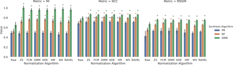
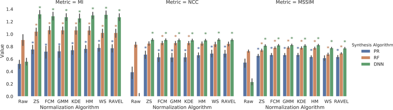
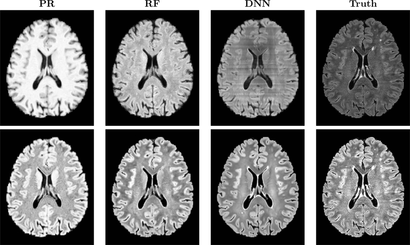
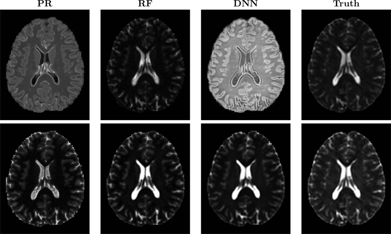
Image synthesis learns a transformation from the intensity features of an input image to yield a different tissue contrast of the output image. This process has been shown to have application in many medical image analysis tasks including imputation, registration, and segmentation. To carry out synthesis, the intensities of the input images are typically scaled-i.e., normalized-both in training to learn the transformation and in testing when applying the transformation, but it is not presently known what type of input scaling is optimal. In this paper, we consider seven different intensity normalization algorithms and three different synthesis methods to evaluate the impact of normalization. Our experiments demonstrate that intensity normalization as a preprocessing step improves the synthesis results across all investigated synthesis algorithms. Furthermore, we show evidence that suggests intensity normalization is vital for successful deep learning-based MR image synthesis.

Keywords: brain MRI; image synthesis; intensity normalization.


Figures

Figure 1.
Figure 1.. T1-to-FLAIR Quality Metrics:
Raw corresponds to synthesis using unnormalized images, ZS to Z-score normalized images, and WS to WhiteStripe normalized images. Statistical significance (denoted by *) for each experiment is compared to Raw (p < 0.05). The error bars represent the 95% confidence interval.
Figure 2.
Figure 2.. T1-to-T2 Quality Metrics:
Raw corresponds to synthesis using unnormalized images, ZS to Z-score normalized images, and WS to WhiteStripe normalized images. Statistical significance (denoted by *) for each experiment is compared to Raw (p < 0.05). The error bars represent the 95% confidence interval.
Figure 3.
Figure 3.. T1-to-FLAIR Synthesis results:
Shown are the results of synthesis using unnormalized (top row) and FCM normalized images (bottom row). The unnormalized DNN result represents a failure of image synthesis.
Figure 4.
Figure 4.. T1-to-T2 Synthesis results:
Shown are the results of synthesis using unnormalized (top row) and FCM normalized images (bottom row). The unnormalized DNN result represents a failure of image synthesis.
Figure 5.
Figure 5.. T1-to-FLAIR DNN Synthesis
Shown from left to right are the results of DNN synthesis using FCM normalized images, unnormalized images, and the ground truth.

Similar articles

Cited by

References

    1. Iglesias JE, Konukoglu E, Zikic D, Glocker B, Leemput KV, and Fischl B, “Is synthesizing MRI contrast useful for inter-modality analysis?,” in [MICCAI], (8149), 631–638 (2013). - PMC - PubMed
    1. Huo Y, Xu Z, Bao S, Assad A, Abramson RG, and Landman BA, “Adversarial synthesis learning enables segmentation without target modality ground truth,” in [2018 IEEE 15th International Symposium on Biomedical Imaging (ISBI 2018)], 1217–1220 (April 2018).
    1. Roy S, Carass A, Jog A, Prince JL, and Lee J, “MR to CT registration of brains using image synthesis,” SPIE Medical Imaging 9034 (2014). - PMC - PubMed
    1. Roy S, Butman JA, and Pham DL, “Robust skull stripping using multiple MR image contrasts insensitive to pathology,” NeuroImage 146, 132–147 (2017). - PMC - PubMed
    1. Roy S, Carass A, and Prince JL, “Magnetic resonance image example-based contrast synthesis,” IEEE Transactions on Medical Imaging 32(12), 2348–2363 (2013). - PMC - PubMed

LinkOut - more resources