Review
. 2019 Aug 27:12:204.
doi: 10.3389/fnmol.2019.00204. eCollection 2019.

New Insights Into Cholinergic Neuron Diversity

Affiliations
Review

New Insights Into Cholinergic Neuron Diversity

Noorya Yasmin Ahmed et al. Front Mol Neurosci. .

Abstract

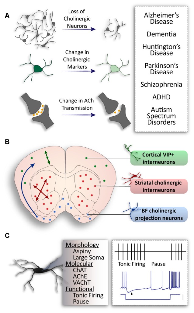
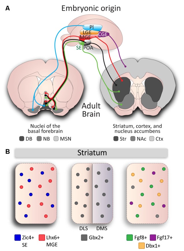
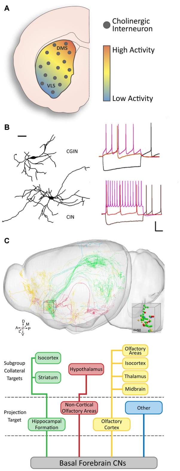
Cholinergic neurons comprise a small population of cells in the striatum but have fundamental roles in fine tuning brain function, and in the etiology of neurological and psychiatric disorders such as Parkinson's disease (PD) or schizophrenia. The process of developmental cell specification underlying neuronal identity and function is an area of great current interest. There has been significant progress in identifying the developmental origins, commonalities in molecular markers, and physiological properties of the cholinergic neurons. Currently, we are aware of a number of key factors that promote cholinergic fate during development. However, the extent of cholinergic cell diversity is still largely underestimated. New insights into the biological basis of their specification indicate that cholinergic neurons may be far more diverse than previously thought. This review article, highlights the physiological features and the synaptic properties that segregate cholinergic cell subtypes. It provides an accurate picture of cholinergic cell diversity underlying their organization and function in neuronal networks. This review article, also discusses current challenges in deciphering the logic of the cholinergic cell heterogeneity that plays a fundamental role in the control of neural processes in health and disease.

Keywords: acetylcholine; development; diversity; interneurons; striatum.


Figures

Figure 1
Figure 1
General populations of cholinergic neurons and alterations in disease. (A) Pathologies associated with cholinergic alteration. (B) Three major populations of cholinergic neurons in the brain. Basal forebrain neurons project to higher structures (blue); striatal interneurons project locally throughout the striatum (red); cortical interneurons co-expressing Vasoactive Intestinal Peptide (VIP) and Choline Acetyltransferase (ChAT) project within cortical layers (green). (C) Common properties of cholinergic neurons: aspiny dendrites and a large soma; expression of ChAT, Acetylcholine Esterase (AChE), Vesicular Acetylcholine Transporter (VAChT) expression; tonic firing patterns with a distinct pause in vivo, and a sag in membrane voltage (arrow) in response to hyperpolarizing current (scale: 100 pA).
Figure 2
Figure 2
Developmental diversity in cholinergic neurons. (A) Cholinergic neurons are derived from different embryonic structures. Proliferative zones of an E13.5 mouse brain embryo: Pallium (Pl), Medial Ganglionic Eminence (MGE), Lateral Ganglionic Eminence (LGE), Septal Epithelium (SE), Preoptic Area (POA), and Caudal Ganglionic Eminence (CGE) give rise to cholinergic neurons of different nuclei: Striatum (Str), Cerebral Cortex (Ctx), Nucleus Accumbens (NAc), and the Basal Forebrain: Nucleus Basalis (NB), Medial Septal Nucleus (MSN), Diagonal Band (DB). (B) Genetic diversity in cholinergic interneurons (CINs) in the striatum. Subsets of CINs express Zic4 (blue) and originate from the SE, and Lhx6 (red) and originate from the MGE. Gbx2 is expressed in almost all CINs and preferentially controls development of late-born CINs in the dorsomedial striatum (DMS), but not in early-born CINs in the dorsolateral striatum (DLS). MGE-derived Fgf8 (green) and Fgf17 (purple), and POA-derived Dbx1 (orange) mark three subgroups defined by genetic expression and reliant on developmental origin, but do not account for the entire CIN population (gray).
Figure 3
Figure 3
Functional diversity of cholinergic neurons. (A) There is a gradient of baseline activity in striatal CINs along the DMS to ventrolateral (VLS) axis (modified from Matamales et al., with permission). (B) Two subsets of striatal CINs show morphological and electrophysiological differences. GABAergic CINs (GCIN) have smaller dendritic fields and slower firing rates than purely cholinergic CINs (adapted from Lozovaya et al., with permission). (C) Basal forebrain cholinergic neurons (CNs) have diverse projection targets and can be grouped into four major categories with subcategories based on collateral targets (adapted from Li et al., with permission).

References

    1. Abudukeyoumu N., Hernandez-Flores T., Garcia-Munoz M., Arbuthnott G. W. (2019). Cholinergic modulation of striatal microcircuits. Eur. J. Neurosci. 49, 604–622. 10.1111/ejn.13949 - DOI - PMC - PubMed
    1. Albert-Gascó H., García-Avilés A., Moustafa S., Sánchez-Sarasua S., Gundlach A. L., Olucha-Bordonau F. E., et al. . (2017). Central relaxin-3 receptor (RXFP3) activation increases ERK phosphorylation in septal cholinergic neurons and impairs spatial working memory. Brain Struct. Funct. 222, 449–463. 10.1007/s00429-016-1227-8 - DOI - PubMed
    1. Allaway K. C., Machold R. (2017). Developmental specification of forebrain cholinergic neurons. Dev. Biol. 421, 1–7. 10.1016/j.ydbio.2016.11.007 - DOI - PubMed
    1. Aoki S., Liu A. W., Akamine Y., Zucca A., Zucca S., Wickens J. R. (2018). Cholinergic interneurons in the rat striatum modulate substitution of habits. Eur. J. Neurosci. 47, 1194–1205. 10.1111/ejn.13820 - DOI - PMC - PubMed
    1. Aoki S., Liu A. W., Zucca A., Zucca S., Wickens J. R. (2015). Role of striatal cholinergic interneurons in set-shifting in the rat. J. Neurosci. 35, 9424–9431. 10.1523/JNEUROSCI.0490-15.2015 - DOI - PMC - PubMed

LinkOut - more resources