

Pleiotropic Roles of P2X7 in the Central Nervous System
- PMID: 31551714
- PMCID: PMC6738027
- DOI: 10.3389/fncel.2019.00401
Pleiotropic Roles of P2X7 in the Central Nervous System
Abstract
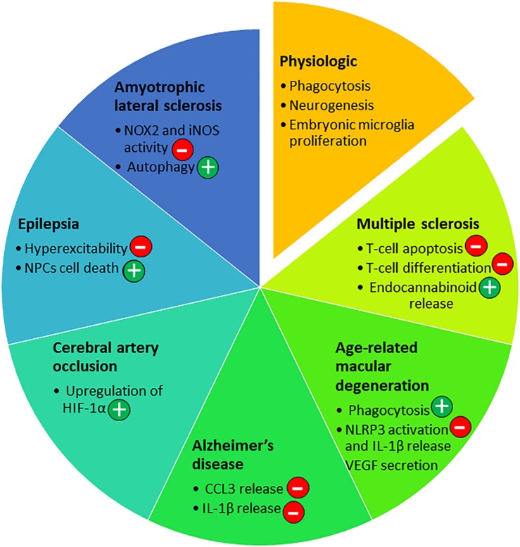
The purinergic receptor P2X7 is expressed in neural and immune cells known to be involved in neurological diseases. Its ligand, ATP, is a signaling molecule that can act as a neurotransmitter in physiological conditions or as a danger signal when released in high amount by damaged/dying cells or activated glial cells. Thus, ATP is a danger-associated molecular pattern. Binding of ATP by P2X7 leads to the activation of different biochemical pathways, depending on the physiological or pathological environment. The aim of this review is to discuss various functions of P2X7 in the immune and central nervous systems. We present evidence that P2X7 may have a detrimental or beneficial role in the nervous system, in the context of neurological pathologies: epilepsy, Alzheimer's disease, multiple sclerosis, amyotrophic lateral sclerosis, age-related macular degeneration and cerebral artery occlusion.
Keywords: ATP; P2X7; animal model; demyelinating disease; nervous system; neurodegenerative disease; neurologic disease; purinergic receptor.
Figures
Similar articles
-
The P2X7 Receptor in Inflammatory Diseases: Angel or Demon?Front Pharmacol. 2018 Feb 6;9:52. doi: 10.3389/fphar.2018.00052. eCollection 2018. Front Pharmacol. 2018. PMID: 29467654 Free PMC article. Review.
-
P2X7-dependent immune pathways in retinal diseases.Neuropharmacology. 2023 Feb 1;223:109332. doi: 10.1016/j.neuropharm.2022.109332. Epub 2022 Nov 11. Neuropharmacology. 2023. PMID: 36372269 Review.
-
Regulation of P2X7 receptor expression and function in the brain.Brain Res Bull. 2019 Sep;151:153-163. doi: 10.1016/j.brainresbull.2018.12.008. Epub 2018 Dec 26. Brain Res Bull. 2019. PMID: 30593878 Review.
-
P2X7-like receptor activation in astrocytes increases chemokine monocyte chemoattractant protein-1 expression via mitogen-activated protein kinase.J Neurosci. 2001 Sep 15;21(18):7135-42. doi: 10.1523/JNEUROSCI.21-18-07135.2001. J Neurosci. 2001. PMID: 11549724 Free PMC article.
-
P2X7 receptor: an emerging target in central nervous system diseases.Trends Pharmacol Sci. 2014 Oct;35(10):537-47. doi: 10.1016/j.tips.2014.08.002. Epub 2014 Sep 12. Trends Pharmacol Sci. 2014. PMID: 25223574 Review.
Cited by
-
Modulation of Microglial Function by ATP-Gated P2X7 Receptors: Studies in Rat, Mice and Human.Cells. 2024 Jan 16;13(2):161. doi: 10.3390/cells13020161. Cells. 2024. PMID: 38247852 Free PMC article. Review.
-
Does Cholinergic Stimulation Affect the P2X7 Receptor-Mediated Dye Uptake in Mast Cells and Macrophages?Front Cell Neurosci. 2020 Oct 28;14:548376. doi: 10.3389/fncel.2020.548376. eCollection 2020. Front Cell Neurosci. 2020. PMID: 33328886 Free PMC article.
-
The P2X7 Receptor, a Multifaceted Receptor in Alzheimer's Disease.Int J Mol Sci. 2023 Jul 21;24(14):11747. doi: 10.3390/ijms241411747. Int J Mol Sci. 2023. PMID: 37511507 Free PMC article. Review.
-
The Role of Cannabinoids in CNS Development: Focus on Proliferation and Cell Death.Cell Mol Neurobiol. 2023 May;43(4):1469-1485. doi: 10.1007/s10571-022-01263-y. Epub 2022 Aug 4. Cell Mol Neurobiol. 2023. PMID: 35925507 Free PMC article. Review.
-
Recombinant Analogs of Sea Anemone Kunitz-Type Peptides Influence P2X7 Receptor Activity in Neuro-2a Cells.Mar Drugs. 2023 Mar 20;21(3):192. doi: 10.3390/md21030192. Mar Drugs. 2023. PMID: 36976241 Free PMC article.
References
-
- Adinolfi E., Callegari M. G., Cirillo M., Pinton P., Giorgi C., Cavagna D., et al. (2009). Expression of the P2X7 receptor increases the Ca2+ content of the endoplasmic reticulum, activates NFATc1, and protects from apoptosis. J. Biol. Chem. 284 10120–10128. 10.1074/jbc.M805805200 - DOI - PMC - PubMed
-
- Adinolfi E., Callegari M. G., Ferrari D., Bolognesi C., Minelli M., Wieckowski M. R., et al. (2005). Basal activation of the P2X7 ATP receptor elevates mitochondrial calcium and potential, increases cellular ATP levels, and promotes serum-independent growth. Mol. Biol. Cell 16 3260–3272. 10.1091/mbc.e04-11-1025 - DOI - PMC - PubMed
Publication types
LinkOut - more resources
Full Text Sources