. 2019 Sep 4:10:967.
doi: 10.3389/fphar.2019.00967. eCollection 2019.

Hydroxycamptothecin Inhibits Peritendinous Adhesion via the Endoplasmic Reticulum Stress-Dependent Apoptosis

Affiliations

Hydroxycamptothecin Inhibits Peritendinous Adhesion via the Endoplasmic Reticulum Stress-Dependent Apoptosis

Zhixiao Yao et al. Front Pharmacol. .

Abstract

Traumatic peritendinous fibrosis is a worldwide clinical problem resulting in severe limb disability. Hydroxycamptothecin (HCPT) is an anti-neoplastic drug widely exploited in clinical practice. It has shown potential of anti-fibrosis in recent years. We previously demonstrated that HCPT inhibited the characterization of fibrosis in vitro. However, it is still unclear whether it ameliorates peritendinous adhesion in an in vivo animal tendon injury model. The underlying mechanism is also worth investigating. The present study aims to determine whether HCPT inhibits tendon adhesion and to explore the underlying mechanisms. In a rat tendon injury model, we observed that topical application of HCPT significantly attenuated peritendinous adhesion as revealed by the results of macroscopic observation, biomechanical, histological, immunohistochemical evaluation, western blot, and quantitative PCR (q-PCR) analyses. Furthermore, western blot and q-PCR analyses revealed that this phenomenon is correlated with HCPT activation of endoplasmic reticulum (ER) stress. In addition, in vitro studies show that HCPT significantly inhibits fibroblast proliferation and induces apoptosis by reducing the expression of extracellular matrix (ECM) proteins COL3A1 and α-smooth muscle actin (α-SMA). Finally, we employed small interfering RNA (siRNA) to target inositol requiring kinase 1 (IRE1) and activated transcription factor 6 (ATF-6) to verify that the effect of inhibitory fibrosis of HCPT disappears after knockdown of ATF-6 and IRE1, thereby suggesting that an anti-fibrotic effect of HCPT is mediated by the ER-dependent apoptotic pathway. In conclusion, our results indicate that HCPT inhibits peritendinous fibrosis through the ER-dependent apoptotic pathway and might serve as a potential solution to prevent traumatic peritendinous adhesion.

Keywords: ATF-6; IRE1; TGF-β1; hydroxycamptothecin; peritendinous fibrosis.


Figures
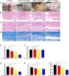
Figure 1
Figure 1
HCPT alleviated peritendinous adhesion in tenon-injured rats. Different concentrations of HCPT (0, 0.01, 0.05, and 0.1 mg/ml) were applied locally to the repaired tendon site for 5 min, and the tendons were examined 3 weeks later. (A) Macroscopic evaluation of peritendinous adhesions. (B-D) Histological assessment of tendon adhesions. (E) Macroscopic adhesion score. (F) Maximum tensile strength. (G, H) Histological score and tissue healing score. (I) Hydroxyproline content. Black arrow indicates adhesion tissue. T: tendon. Data are expressed as the mean ± SD of five independent samples. *P < 0.05. ns: not significant.
Figure 2
Figure 2
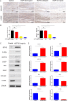
HCPT inhibited adhesion and activated ER stress in vivo. Different concentrations of HCPT (0, 0.01, 0.05, and 0.1 mg/ml) were applied locally to the tendon surgery site for 5 min, and the tendon was collected 3 weeks later. (A) Representative images of the immunohistochemical staining of COL3A1 and α-SMA. (B) Relative levels of COL3A1 and α-SMA. Tendons were treated with or without HCPT and collected 3 weeks after surgery for testing. (C) Representative immunoblot images of ATF-6, P-IRE1, GRP78, CHOP, Bax, Bal-2, COL3A1, and α-SMA. (D) Q-PCR analysis of the mRNA expression levels of ATF-6, IRE1, GRP78, CHOP, Bax, Bal-2, COL3A1 and α-SMA. Data are showed as the mean ± SD of five independent samples. *P < 0.05. ns: not significant.
Figure 3
Figure 3
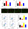
Effects of HCPT on the proliferation and apoptosis of fibroblasts. Fibroblasts were incubated with TGF-β1 (2 ng/ml) in the absence or presence of HCPT (1 μg/ml) for 24 h. (A) CCK-8 assay of cell viability. (B) Quantitative analysis of EdU staining. (C) Representative images of dead/live staining and (E) dead/live rate analysis. (D) Representative pictures of apoptotic flow cytometry and (F) quantitative analysis of fibroblasts apoptosis. Data are expressed as the mean ± SD of five independent samples. *P < 0.05.
Figure 4
Figure 4
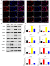
HCPT inhibited TGF-β1-induced adhesion in vitro. Fibroblasts were incubated with HCPT and/or TGF-β1 for 24 h. (A) Representative cellular immunofluorescence pictures of COL3A1 and α-SMA. (B) Representative western blot pictures of ATF-6, P-IRE1, GRP78, CHOP, Bax, Bal-2, COL3A1, and α-SMA. (C) Relative mRNA expression levels of ATF-6, IRE1, GRP78, CHOP, Bax, Bal-2, COL3A1, and α-SMA. Data are showed as the mean ± SD of five independent samples. *P < 0.05. ns: not significant.
Figure 5
Figure 5
SiRNA targeting IRE1 abolished the inhibitory effect of HCPT on TGF-β1-induced fibroblast fibrosis. Fibroblasts were pretreated with siRNA targeting IRE1 and then incubated with TGF-β1 and/or HCPT for 24 h. (A) Representative western blot images of IRE1. (B) The CCK-8 assay of cell viability. (D) Representative pictures of dead/live staining and (C) dead/live rate analysis. (E) Representative cellular immunofluorescence pictures of COL3A1 and α-SMA. (F) Representative western blot pictures of GRP78, CHOP, Bax, Bal-2, COL3A1, and α-SMA. (G) Relative mRNA levels of GRP78, CHOP, Bax, Bal-2, COL3A1, and α-SMA. Data are showed as the mean ± SD of five independent samples. *P < 0.05. ns: not significant.
Figure 6
Figure 6
SiRNA targeting ATF-6 blocked the inhibitory effect of HCPT on TGF-β1-induced fibroblast fibrosis. Fibroblasts were pretreated with siRNA targeting ATF-6 then incubated with TGF-β1 and/or HCPT for 24 h. (A) Representative western blot image of ATF-6. (B) The CCK-8 assay of cell viability. (D) Representative pictures of dead/live staining and (C) dead/live rate analysis. (E) Representative cellular immunofluorescence pictures of COL3A1 and α-SMA. (F) Representative western blot pictures of GRP78, CHOP, Bax, Bal-2, COL3A1, and α-SMA. (G) Relative mRNA expression levels of GRP78, CHOP, Bax, Bal-2, COL3A1, and α-SMA. Data are showed as the mean ± SD of five independent samples. *P < 0.05. ns: not significant.
Figure 7
Figure 7
Potential signaling pathways involved in the regulation of peritendinous adhesion by HCPT.

References

    1. Adachi Y., Yamamoto K., Okada T., Yoshida H., Harada A., Mori K. (2008). ATF6 is a transcription factor specializing in the regulation of quality control proteins in the endoplasmic reticulum. Cell Struct. Funct. 33 (1), 75–89. 10.1247/csf.07044 - DOI - PubMed
    1. Binet F., Sapieha P. (2015). ER stress and angiogenesis. Cell Metab. 22 (4), 560–575. 10.1016/j.cmet.2015.07.010 - DOI - PubMed
    1. Chen S., Jiang S., Zheng W., Tu B., Liu S., Ruan H., et al. (2017). RelA/p65 inhibition prevents tendon adhesion by modulating inflammation, cell proliferation, and apoptosis. Cell Death Dis. 8 (3), e2710. 10.1038/cddis.2017.135 - DOI - PMC - PubMed
    1. Chern Y. J., Wong J. C. T., Cheng G. S. W., Yu A., Yin Y., Schaeffer D. F., et al. (2019). The interaction between SPARC and GRP78 interferes with ER stress signaling and potentiates apoptosis via PERK/eIF2α and IRE1α/XBP-1 in colorectal cancer. Cell Death Dis. 10 (7), 504. 10.1038/s41419-019-1687-x - DOI - PMC - PubMed
    1. Cianfarani F., De Domenico E., Nystrom A., Mastroeni S., Abeni D., Baldini E., et al. (2018). Decorin counteracts disease progression in mice with recessive dystrophic epidermolysis bullosa. Matrix Biol. 81, 3–16. 10.1016/j.matbio.2018.12.001 - DOI - PubMed