. 2019 Sep 10:10:988.
doi: 10.3389/fphar.2019.00988. eCollection 2019.

Erxian Decoction Attenuates TNF-α Induced Osteoblast Apoptosis by Modulating the Akt/Nrf2/HO-1 Signaling Pathway

Affiliations

Erxian Decoction Attenuates TNF-α Induced Osteoblast Apoptosis by Modulating the Akt/Nrf2/HO-1 Signaling Pathway

Nani Wang et al. Front Pharmacol. .

Abstract

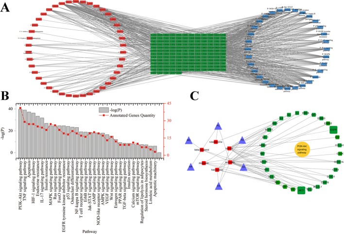
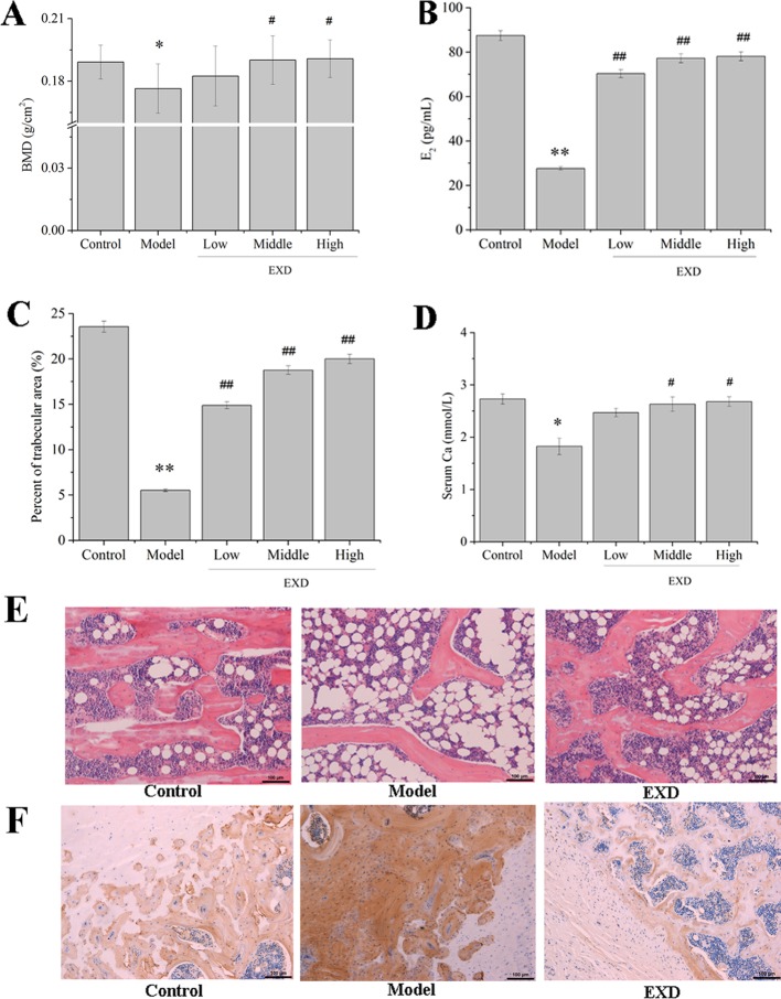
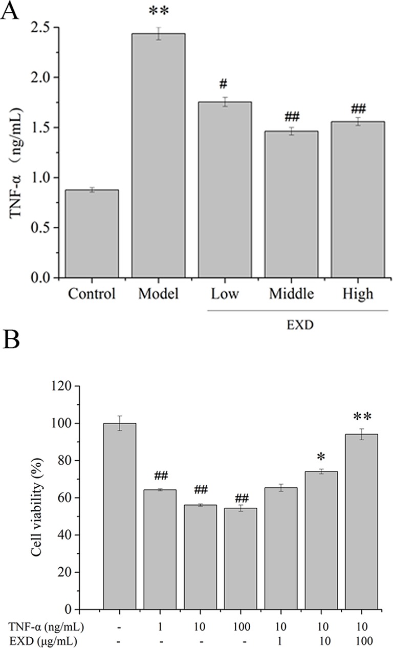
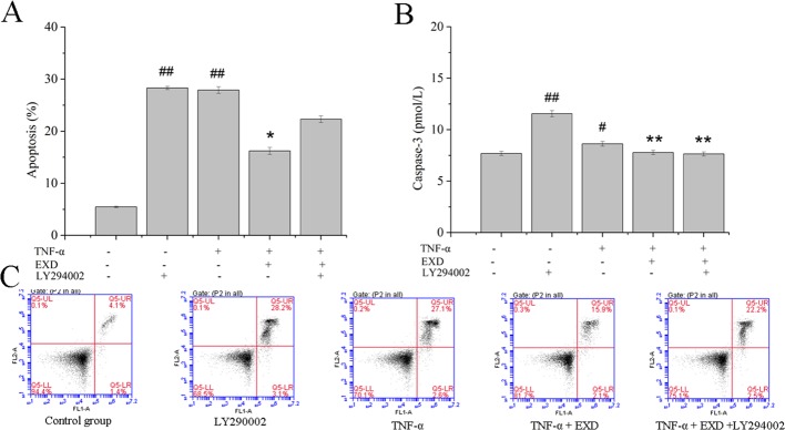
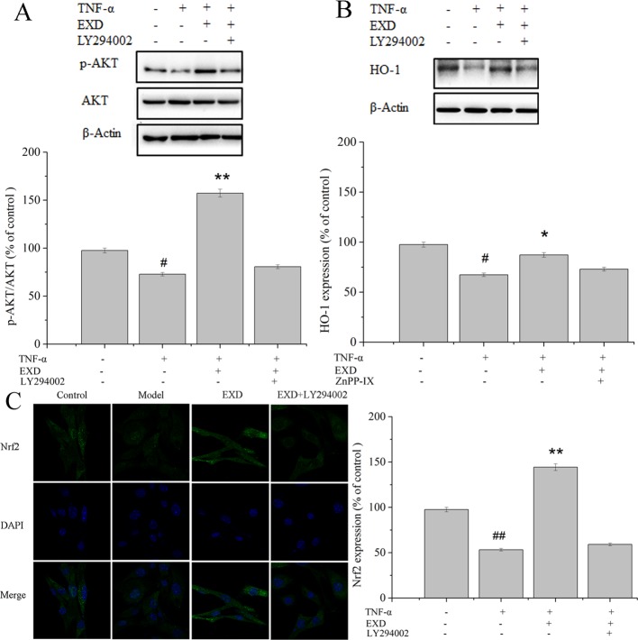
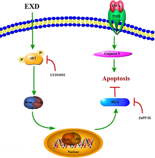
Erxian decoction (EXD), a traditional Chinese medicine formula, has been used for treatment of osteoporosis for many years. The purpose of this study was to investigate the pharmacological effect of EXD in preventing osteoblast apoptosis and the underlying mechanism of prevention. Putative targets of EXD were predicted by network pharmacology, and functional and pathway enrichment analyses were also performed. Evaluations of bone mineral density, serum estradiol level, trabecular area fraction, serum calcium levels, and tumor necrosis factor (TNF)-α levels in ovariectomized rats, as well as cell proliferation assays, apoptosis assays, and western blotting in MC3T3-E1 osteoblasts were performed for further experimental validation. Ninety-three active ingredients in the EXD formula and 259 potential targets were identified. Functional and pathway enrichment analyses indicated that EXD significantly influenced the PI3K-Akt signaling pathway. In vivo experiments indicated that EXD treatment attenuated bone loss and decreased TNF-α levels in rats with osteoporosis. In vitro experiments showed that EXD treatment increased cell viability markedly and decreased levels of caspase-3 and the rate of apoptosis. It also promoted phosphorylation of Akt, nuclear translocation of transcription factor NF-erythroid 2-related factor (Nrf2), and hemeoxygenase-1 (HO-1) expression in TNF-α-induced MC3T3-E1 cells. Our results suggest that EXD exerted profound anti-osteoporosis effects, at least partially by reducing production of TNF-α and attenuating osteoblast apoptosis via Akt/Nrf2/HO-1 signaling pathway.

Keywords: Akt; Erxian decoction; network pharmacology; osteoporosis; tumor necrosis factor.


Figures

Figure 1
Figure 1
(A) Potential active ingredient-target-pathways network of EXD acting on osteoporosis. The network was based on the interaction among ingredients in EXD, active components, and osteoporosis-related targets. Red nodes represented the ingredients; green nodes represented the targets; blue nodes represented the osteoporosis-related pathways. (B) KEGG pathways for potential targets of Erxian decoction. (C) The interaction among herbs, components, and targets in PI3K-Akt signaling pathway.
Figure 2
Figure 2
EXD ameliorated osteoporosis in OVX rats. Wistar rats were administrated with water or EXD extract at a dose of 2 g/kg/day (low level), 4 g/kg/day (middle level), 6 g/kg/day (high level), which started on day 4 after OVX operation for 12 weeks. (A) The bone mineral density. (B) Serum estradiol level. (C) Percent of trabecular area. (D) Serum Ca level. (E) Representative images of femur stained with hematoxylin–eosin (magnification, ×200). Oral administration dose of EXD was 4 g/kg/day. (F) Representative images of femur stained with osteocalcin antibody (magnification, ×200). Oral administration dose of EXD was 4 g/kg/day. #P < 0.05, ##P < 0.01 compared to the control group. *P < 0.05, **P < 0.01 compared to the model group.
Figure 3
Figure 3
(A) TNF-α level in ovariectomy rats. Wistar rats were administrated with water or EXD extract at a dose of 2 g/kg/day (low level), 4 g/kg/day (middle level), 6 g/kg/day (high level), which started on day 4 after OVX operation for 12 weeks. # P <0.05, ## P < 0.01 compared to the control group. *P < 0.05, **P < 0.01 compared to the model group. (B) Effects of EXD on MC3T3-E1cell viability. Cells were induced by TNF-α. #P <0.05, ##P < 0.01 compared to the control group. *P < 0.05, **P < 0.01 compared to the TNF-α group (10 ng/ml).
Figure 4
Figure 4
Effect of EXD and selective inhibitor LY294002 on TNF-α-induced apoptosis in MC3T3-E1 cells. Cells were incubated with or without 20 μM LY294002, 100 μg/ml EXD, and 10 ng/ml TNF-α for 24 h. (A) Cell apoptosis rate. (B) Caspase-3 level. (C) Annexin V/PI staining results. #P < 0.05, ##P < 0.01 compared to the control group. *P < 0.05, **P < 0.01 compared to the TNF-α group.
Figure 5
Figure 5
Effect of EXD and selective inhibitor LY294002 on (A) AKT, (B) HO-1, and (C) Nrf2 expression (magnification, ×650). Cells were incubated with or without 20 μM LY294002, 5 μM ZnPP-IX, 100 μg/ml EXD, and 10 ng/ml TNF-α for 24 h. #P < 0.05, ##P < 0.01 compared to the control group. *P < 0.05, **P < 0.01 compared to the TNF-α group.
Figure 6
Figure 6
A proposed signaling pathway involved in EXD against TNF-α-induced cell damage.

References

    1. Bian Q., Yang H., Chan C. O., Jin D., Mok D. K., Chen S. (2013). Fingerprint analysis and simultaneous determination of phenolic compounds in extracts of Curculiginis Rhizoma by HPLC-diode array detector. Chem. Pharm. Bull. 61 (8), 802–808. 10.1248/cpb.c12-01058 - DOI - PubMed
    1. Bian J., Xu S., Huang S., Wang Z. (1996). A Study on the chemical constituents of Anemarrhena asphodeloides Bge. Shenyang Yao Ke Da Xue Xue Bao 13 (66), 34–40. 10.1248/cpb.c12-01058 - DOI
    1. Cao D., Han T., Zheng Y., Qin L., Zhang Q. (2009). Phenolic glycosides and lignans components in Curculigo orchioides Gaertn. Acad. J. Second Mil. Med. Univ. 29 (2), 194–197. 10.3724/SP.J.1008.2009.00194 - DOI
    1. Chen C. C., Huang Y. L., Sun C. M., Shen C. C. (1996). New prenylflavones from the leaves of Epimedium sagittatum. J. Nat. Prod. 59 (4), 412–414. 10.1021/np9601925 - DOI - PubMed
    1. Chen X., Ji Z. L., Chen Y. Z. (2002). TTD: therapeutic target database. Nucleic Acids Res. 30 (1), 412–415. 10.1093/nar/30.1.412 - DOI - PMC - PubMed