Review
. 2019 Sep 10:10:970.
doi: 10.3389/fneur.2019.00970. eCollection 2019.

Architecture of the Cutaneous Autonomic Nervous System

Affiliations
Review

Architecture of the Cutaneous Autonomic Nervous System

Patrick Glatte et al. Front Neurol. .

Abstract

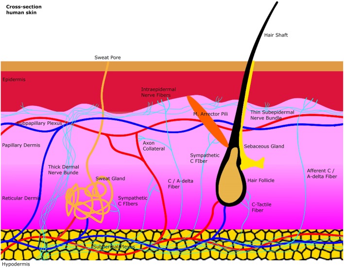
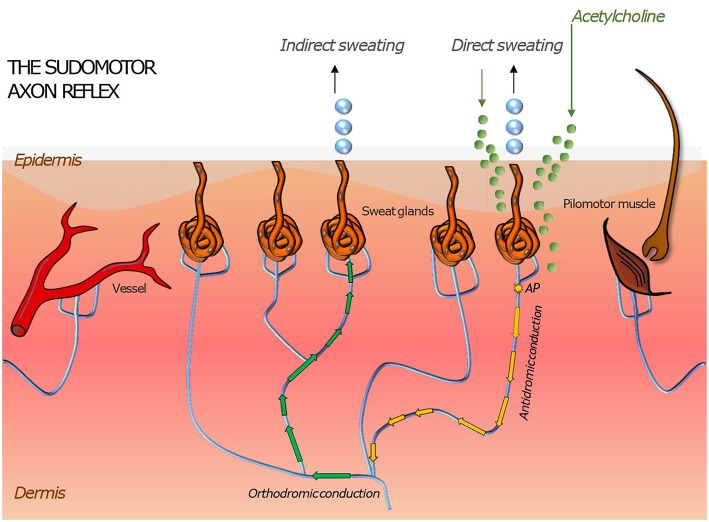
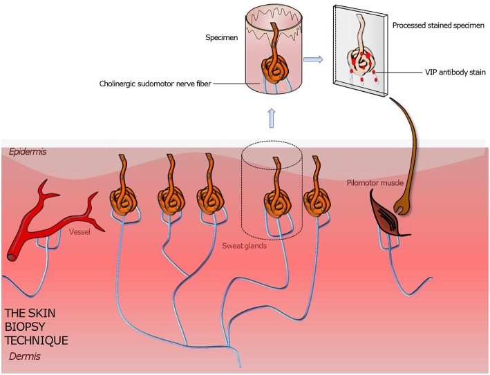
The human skin is a highly specialized organ for receiving sensory information but also to preserve the body's homeostasis. These functions are mediated by cutaneous small nerve fibers which display a complex anatomical architecture and are commonly classified into cutaneous A-beta, A-delta and C-fibers based on their diameter, myelinization, and velocity of conduction of action potentials. Knowledge on structure and function of these nerve fibers is relevant as they are selectively targeted by various autonomic neuropathies such as diabetic neuropathy or Parkinson's disease. Functional integrity of autonomic skin nerve fibers can be assessed by quantitative analysis of cutaneous responses to local pharmacological induction of axon reflex responses which result in dilation of cutaneous vessels, sweating, or piloerection depending on the agent used to stimulate this neurogenic response. Sensory fibers can be assessed using quantitative sensory test. Complementing these functional assessments, immunohistochemical staining of superficial skin biopsies allow analysis of structural integrity of cutaneous nerve fibers, a technique which has gained attention due to its capacity of detecting pathogenic depositions of alpha-synuclein in patients with Parkinson's disease. Here, we reviewed the current literature on the anatomy and functional pathways of the cutaneous autonomic nervous system as well as diagnostic techniques to assess its functional and structural integrity.

Keywords: C-fiber; Parkinson's disease; autonomic (vegetative) nervous system; autonomic neuropathy; axon-reflex; diabetes; punch skin biopsy; skin.


Figures

Figure 1
Figure 1
A simplified illustration of the general anatomy of the skin with the focus on autonomic nerve fibers and their innervated organs. Sweat glands, blood vessels and the arrector pili muscle are innervated by sympathetic C-fibers in the dermis. Afferent intraepidermal nerve fibers of the class C and Aδ are found in the epidermis as free nerve endings. Axon collaterals of these afferent fibers also supply blood vessels with efferent antidromic control. Small sensory fibers branch off from thicker dermal nerve bundles to create thinner subepidermal nerve bundles that innervate the epidermis.
Figure 2
Figure 2
Illustration of skin organs innervated by the autonomic nervous system with an axon reflex mediated in sudomotor nerve fibers by iontophoretic application of acetylcholine to the skin. Following a direct sweat response in the area of acetylcholine application, an action potential travels antidromically and then orthodromically to a neighboring population of sweat glands where it evokes “indirect” sweating in a skin region adjacent to the region of iontophoresis. Similar responses can be induced in pilomotor and vasomotor fibers. Their magnitude is a surrogate measure of functional integrity of the autonomic nerve fiber mediating the axon reflex.
Figure 3
Figure 3
Illustration of a punch skin biopsy on eccrine sweat glands to quantify the cholinergic sudomotor nerve fibers. The specimen is fixed, sectioned, and stained with antibodies for PGP 9,5 (the pan axonal marker), tyrosine hydroxylase (a sweat gland neuroendocrine cell marker), and VIP (a marker for sympathetic nerve fibers) to highlight the sought-after tissue. Further various quantitation methods are applied to assess the sweat gland nerve fiber density. Based on this technique pilomotor and vasomotor autonomic nerve fibers can be quantified by using suitable staining methods. A comparison of the determined nerve fiber density to those of normative datasets gives information about the functionality and condition of the autonomic nervous system innervating skin organs.

References

    1. Peters MJ, Bakkers M, Merkies IS, Hoeijmakers JG, van Raak EP, Faber CG. Incidence and prevalence of small-fiber neuropathy: a survey in the Netherlands. Neurology. (2013) 81:1356–60. 10.1212/WNL.0b013e3182a8236e - DOI - PubMed
    1. Chan AC, Wilder-Smith EP. Small fiber neuropathy: getting bigger! Muscle Nerve. (2016) 53:671–82. 10.1002/mus.25082 - DOI - PubMed
    1. Freeman R. Autonomic peripheral neuropathy. Lancet. (2005) 365:1259–70. 10.1016/S0140-6736(05)74815-7 - DOI - PubMed
    1. Donadio V, Incensi A, Giannoccaro MP, Cortelli P, Stasi VD, Pizza F, et al. Peripheral autonomic neuropathy: diagnostic contribution of skin biopsy. J Neuropathol Exp Neurol. (2012) 71:1000–8. 10.1097/NEN.0b013e3182729fdc - DOI - PubMed
    1. Frerichs KU, Feuerstein GZ. Laser-Doppler flowmetry. A review of its application for measuring cerebral and spinal cord blood flow. Mol Chem Neuropathol. (1990) 12:55–70. 10.1007/BF03160057 - DOI - PubMed