. 2019 Sep 3:10:1942.
doi: 10.3389/fmicb.2019.01942. eCollection 2019.

The Tyrosine-Autokinase UbK Is Required for Proper Cell Growth and Cell Morphology of Streptococcus pneumoniae

Affiliations

The Tyrosine-Autokinase UbK Is Required for Proper Cell Growth and Cell Morphology of Streptococcus pneumoniae

Anaïs Pelletier et al. Front Microbiol. .

Abstract

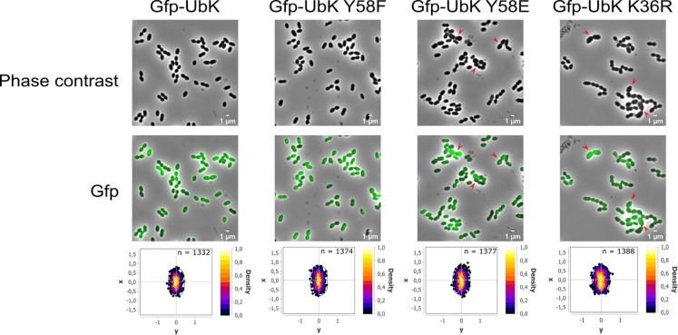
Protein phosphorylation is a key post-translational modification required for many cellular functions of the bacterial cell. Recently, we identified a new protein-kinase, named UbK, in Bacillus subtilis that belongs to a new family of protein-kinases widespread in bacteria. In this study, we analyze the function of UbK in Streptococcus pneumoniae. We show that UbK displays a tyrosine-kinase activity and autophosphorylates on a unique tyrosine in vivo. To get insights into its cellular role, we constructed a set of pneumococcal ubk mutants. Using conventional and electron microscopy, we show that the ubk deficient strain, as well as an ubk catalytic dead mutant, display both severe cell-growth and cell-morphology defects. The same defects are observed with a mutant mimicking permanent phosphorylation of UbK whereas they are not detected for a mutant mimicking defective autophosphorylation of UbK. Moreover, we find that UbK phosphorylation promotes its ability to hydrolyze ATP. These observations show that the hydrolysis of ATP by UbK serves not only for its autophosphorylation but also for a distinct purpose essential for the optimal cell growth and cell-morphogenesis of the pneumococcus. We thus propose a model in which the autophosphorylation/dephosphorylation of UbK regulates its cellular function through a negative feedback loop.

Keywords: ATP hydrolysis; Streptococcus pneumoniae; cell-morphogenesis; protein phosphorylation; tyrosine-kinase.


Figures

FIGURE 1
FIGURE 1
Deletion of ubk and co-selection of a suppressive mutation. (A) Compared transformation frequencies of the WT (clear bars) and the ubk+-ubkCEP (dark bars) recipient strains transformed either with genomic DNA of the Δubk:spc-rpsL original ubk mutant (genomic) or with a PCR overlapping the Δubk:spc-rpsL mutation (PCR). The transformation ratio Δubk DNA/reference gDNA represents the SpcR transformants numbered with Δubk transforming gDNA or Δubk transforming PCR DNA relative to SpcR transformants numbered with the ΔphpP-stkP:spc-rpsL transforming genomic control DNA used as a reference (reference gDNA). (B) Compared transformation frequencies of the WT (clear bars) and the suppressor-WT (dark bars) recipient strains transformed with a PCR overlapping the Δubk:kan-rpsL mutation. The transformation ratio Δubk PCR DNA/reference gDNA represents the KanR transformants numbered with the Δubk:kan-rpsL transforming PCR DNA relative to KanR transformants numbered with the Δspr1424:kan-rpsL transforming genomic control DNA used as a reference (reference gDNA). For (A,B) panels, bars represent standard deviations. Experiments were led in triplicate.
FIGURE 2
FIGURE 2
Autophosphorylation of UbK mutant proteins purified from E. coli or purified directly from S. pneumoniae. (Left) Immunodetection with antiphosphotyrosine antibodies of Tyrosine phosphorylation of UbK and UbK-K36R purified from E. coli. (Middle and right) Immunodetection with antiphosphotyrosine antibodies of Tyrosine-autophosphorylation of GFP-UbK and GFP-UbK mutant proteins directly immuno-purified from S. pneumoniae. (A) Staining with Coomassie blue of GFP-UbK, GFP-UbK-K36R and GFP-UbK-Y58F. (B) Immunodetection of Tyrosine-autophosphorylation on Tyr58 of GFP-UbK. In both cases proteins were analyzed by SDS-PAGE, transferred onto a PVDF membrane and immunodetection of Tyrosine phosphorylation was done with antiphosphotyrosine 4G10 antibodies.
FIGURE 3
FIGURE 3
Representative growth of ubk mutants. Growth curves of ubk mutants at 37°C: WT (black squares), Δubk (white triangles), ubk-K36R (white circles), ubk-Y58E (black circles), ubk-Y58F (black crosses). Bacteria were diluted so that 2.105 cells were added at t = 0 min. ubk-Y58F strain grows like the WT strain whereas Δubk, ubk-K36R and ubk-Y58E strains display an increased lag phase and a reduced generation time during the exponential growth phase. Growths were led in triplicate.
FIGURE 4
FIGURE 4
ATP hydrolytic activity of WT and mutated UbK-6His proteins. (A) Hydrolysis of ATP is monitored during 50 min at 37°C by measuring the disappearance of NADH at 340 nm. Bars are representative of the relative ATP hydrolytic activity. The activity of UbK WT is standardized at 1. Standard deviation is represented by vertical bars. An ANNOVA test was performed using the GraphPad software (****P < 0.0001 and ∗∗P < 0.005). The experiment was repeated three times. (B) Specific ATP hydrolytic activities of the UbK-6His proteins in nmol.mg–1.min–1
FIGURE 5
FIGURE 5
Cell morphology of ubk mutants and cell-width analysis. Phase contrast microscopy micrographs of the WT, Δubk, complemented (ubkCEP−Δubk), ubk-K36R, ubk-Y58F, and ubk-Y58E strains. Arrows show swelled mutant cells. Scale bar, 1 μm. Violin plot showing the distribution of the cell width (μm) for each strain. Mann–Whitney test (****P < 0.0001). The distribution of the cell width of mutants with morphological defects is shown in red. The number of cells scored and analyzed for each strain is indicated. For each violin, the width of the shaded area represents the proportion of cells located there. The bottom and top of the inside-box represent the 25th and 75th percentile. The bar in the box indicates the median value while the black dot indicates the mean value.
FIGURE 6
FIGURE 6
Scanning and transmission electron micrographs of WT strain and ubk mutants. From left to right column: scanning electron micrographs with the lowest power magnifier (scale bar, 1 μm), scanning electron micrographs with the highest power magnifier (scale bar, 100 nm) and transmission electron micrographs (scale bar, 200 nm). Red arrows on scanning electron micrographs show gashes or possible zones of peeling on the cell-wall of the mutants. Red arrows on transmission electron micrographs show zones of peeling on the cell-wall; black arrows show mispositioned septa.
FIGURE 7
FIGURE 7
Cytoplasmic localization of the different GFP-UbK forms. Phase-contrast images (top row) and GFP images (bottom row) of exponentially growing cells expressing GFP-UbK or GFP-UbK-K36R or GFP-UbK-Y58F or GFP-UbK-Y58E. Bar scale 1 μm. Heat maps represent the localization patterns of GFP-UbK or GFP-UbK-K36R or GFP-UbK-Y58F or GFP-UbK-Y58E. The n values represent the number of cells analyzed.
FIGURE 8
FIGURE 8
Model depicting the function of UbK as an ATP hydrolytic enzyme and as a tyrosine-autokinase. For each version of UbK, the ATP hydrolysis activity is shown with an arrow whose size depends on the relative level of this enzymatic activity. WT UbK, mutants of the lysine 36 of the Walker A motif, as well as mutants of the phospho-site (Tyrosine 58) are represented with a “smile” illustrating whether the mutation is beneficial or detrimental for the cell growth and the morphology of S. pneumoniae.

References

    1. Akochy P. M., Bernard D., Roy P. H., Lapointe J. (2004). Direct glutaminyl-tRNA biosynthesis and indirect asparaginyl-tRNA biosynthesis in Pseudomonas aeruginosa PAO1. J. Bacteriol. 186 767–776. 10.1128/jb.186.3.767-776.2004 - DOI - PMC - PubMed
    1. Beilharz K., Novakova L., Fadda D., Branny P., Massidda O., Veening J. W. (2012). Control of cell division in Streptococcus pneumoniae by the conserved Ser/Thr protein kinase StkP. Proc. Natl. Acad. Sci. U.S.A. 109 E905–E913. 10.1073/pnas.1119172109 - DOI - PMC - PubMed
    1. Berge M. J., Mercy C., Mortier-Barriere I., Vannieuwenhze M. S., Brun Y. V., Grangeasse C., et al. (2017). A programmed cell division delay preserves genome integrity during natural genetic transformation in Streptococcus pneumoniae. Nat. Commun. 8:1621. 10.1038/s41467-017-01716-9 - DOI - PMC - PubMed
    1. Bidnenko V., Shi L., Kobir A., Ventroux M., Pigeonneau N., Henry C., et al. (2013). Bacillus subtilis serine/threonine protein kinase YabT is involved in spore development via phosphorylation of a bacterial recombinase. Mol. Microbiol. 88 921–935. 10.1111/mmi.12233 - DOI - PMC - PubMed
    1. Borchert N., Dieterich C., Krug K., Schutz W., Jung S., Nordheim A., et al. (2010). Proteogenomics of Pristionchus pacificus reveals distinct proteome structure of nematode models. Genome Res. 20 837–846. 10.1101/gr.103119.109 - DOI - PMC - PubMed

LinkOut - more resources