. 2019 Sep 6:10:2043.
doi: 10.3389/fmicb.2019.02043. eCollection 2019.

SR-BI Interactome Analysis Reveals a Proviral Role for UGGT1 in Hepatitis C Virus Entry

Affiliations

SR-BI Interactome Analysis Reveals a Proviral Role for UGGT1 in Hepatitis C Virus Entry

Jiazhao Huang et al. Front Microbiol. .

Abstract

Hepatitis C virus (HCV) entry is mediated by multiple co-receptors including scavenger receptor class B, type I (SR-BI). To elucidate the interactome of human SR-BI, we performed immunoprecipitation (IP) experiment coupled with mass spectrometry (MS) analysis. UDP-glucose:glycoprotein glucosyltransferase 1 (UGGT1), a key component of calnexin cycle involved in protein glycosylation, was identified as a SR-BI-interacting protein. Silencing UGGT1 or N-glycosylation inhibitor treatment reduced SR-BI protein level. Further study demonstrated that human SR-BI was N-glycosylated at nine asparagines. Moreover, HCV entry and infection were reduced by the absence of UGGT1. Interestingly, silencing SR-BI reduced protein stability of UGGT1 and protein quality control function mediated by UGGT1. Our finding not only identified UGGT1 as a HCV host factor, but also identified a UGGT1-mediated protein folding function for SR-BI.

Keywords: HCV; N-glycosylation; SR-BI; UGGT1; calnexin.


Figures

FIGURE 1
FIGURE 1
Interactome analysis of SR-BI. (A) String analysis showed top 10 proteins that interacted with SR-BI from LC-MS/MS experiments. (B–D) Reactome Pathways (B), Biological Process (C) and KEGG Pathway (D) analysis of the top 10 potential binding partners of SR-BI.
FIGURE 2
FIGURE 2
Validation of the interactions between SR-BI and components of calnexin cycle. (A) Co-immunoprecipitation assay showed SR-BI-Flag could interact with UGGT1 and calnexin. SR-BI-Flag or p3xFlag-CMV-14 empty construct was transfected into 293T cells for 48 h. The cell lysates were incubated with anti-Flag antibody-coated beads and co-IP samples were subjected to SDS-PAGE followed by Western blotting with indicated antibodies. marked antibody heavy chain. (B) UGGT1 interacted with SR-BI but not other HCV receptors. Four constructs expressing C-termed Flag tagged HCV receptors or p3xFlag-CMV-14 empty vectors were transfected into 293T cells for 48 h. Cell lysates were incubated with anti-Flag antibody-coated beads and detected by SDS-PAGE followed by Western blotting with indicated antibodies. (C) Co-immunoprecipitation assay showed calnexin interacted with SR-BI and UGGT1. Calnexin-Flag construct was transfected into 293T cells for 48 h and then cell lysates were incubated with anti-Flag antibody or control mouse IgG -coated beads. Co-IP samples were subjected to SDS-PAGE followed by Western blotting with indicated antibodies. (D) Co-immunoprecipitation assay of endogenous SR-BI showed SR-BI interacted with UGGT1 and calnexin. Huh7.5.1 cell lysates were incubated with anti-SR-BI antibody or control mouse IgG-coated beads. Co-IP samples were subjected to SDS-PAGE followed by Western blotting with indicated antibodies.
FIGURE 3
FIGURE 3
Scavenger receptor class B, type I partially colocalized with UGGT1 and calnexin. (A) Colocalization of SR-BI with calnexin-Flag in Huh7.5.1 cells. Huh7.5.1 cells transfected with calnexin-Flag construct were fixed by methanol, permealized by 0.5% Triton X-100 and immunofluorescently labeled for SR-BI antibody (green) and Flag antibody (red). DAPI marked nucleus (blue). Scale bar, 15 μm. (B) Colocalization of SR-BI with UGGT1-Flag in Huh7.5.1 cells. Huh7.5.1 cells transfected with UGGT1-Flag construct were fixed by methanol, permealized by 0.5% Triton X-100 and immunofluorescently labeled for SR-BI antibody (green) and Flag antibody (red). DAPI marked nucleus (blue). Scale bar, 15 μm.
FIGURE 4
FIGURE 4
Silencing UGGT1 or tunicamycin treatment reduced protein level of SR-BI. (A) Silencing UGGT1 reduced SR-BI protein quantity. Huh7.5.1 cells were treated with UGGT1 siRNA#1or control siRNA for 72 h and then cell lysates were subjected to SDS-PAGE followed by Western blotting with indicated antibodies. The blots of three independent experiments were quantified using ImageJ. Relative protein level of SR-BI was normalized to the blot signal of actin. (B) Huh7.5.1 cells were treated with siRNA targeting UGGT1 or control siRNA for 72 h. Total RNA was isolated and reverse transcribed and then the qPCR was performed. Relative SR-BI mRNA level was normalized to GAPDH mRNA level. (C) Protein level of SR-BI was decreased by tunicamycin, an inhibitor of N-glycosylation. Huh7.5.1 cells were treated with different dosages of tunicamycin for 16 h and then the cell lysates were subjected to SDS-PAGE followed by Western blotting with indicated antibodies. (D) PNGase F reduced the size of SR-BI-Flag. Huh7.5.1 cells transfected with SR-BI construct were treated with PNGase F or mock treatment. Then cell lysates were subjected to SDS-PAGE followed by Western blotting with indicated antibodies. (E) Size of endogenous SR-BI was decreased by PNGase F. Cell lysates from Huh7.5.1 cells treated with PNGase F or mock treatment were subjected to SDS-PAGE followed by Western blotting with indicated antibodies. ∗∗∗P < 0.001.
FIGURE 5
FIGURE 5
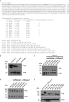
Human SR-BI was N-glycosylated. (A) Prediction of potential N-glycosylation site in human SR-BI. Human SR-BI sequence was predicted through N-Gly website (http://www.cbs.dtu.dk/services/NetNGlyc/). The potential N-glycosylation sites of human SR-BI was labeled in red. SR-BI constructs with N to Q or D mutation were shown. (B) SR-BI was decreased when four N were mutated to Q(N4Q). 293T cells were transfected with constructs expressing Flag tagged SR-BI-WT, SR-BI N4Q, SR-BI N9Q for 48 h and then cell lysates were subjected to SDS-PAGE followed by Western blotting with indicated antibodies. (C) Size of SR-BI of N4Q was reduced by PNGase F in the presence of MG132. 293T cells were transfected with constructs expressing Flag tagged SR-BI-WT, SR-BI N4Q, SR-BI N9Q for 48 h and MG132 was presence in the last 12 h. Cell lysates were treated with PNGase F or mock treatment and then subjected to SDS-PAGE followed by Western blotting with indicated antibodies. (D) Size of SR-BI of N2Q mutant was between N5Q and WT while PNGase F decreased all of them to the same size. 293T cells were transfected with constructs expressing Flag tagged SR-BI-WT, SR-BI N2Q, or SR-BI N5Q for 48 h and cell lysates were treated with PNGase F or mock treatment. The samples were subjected to SDS-PAGE followed by Western blotting with indicated antibodies. (E) Size of SR-BI became smaller with increased mutation sites. 293T cells were transfected with constructs expressing Flag tagged SR-BI-WT, SR-BI N2Q, SR-BI N4Q, SR-BI N5Q, SR-BI N6Q, SR-BI N7Q, or SR-BI N9D for 48 h and cell lysates were subjected to SDS-PAGE followed by Western blotting with indicated antibodies.
FIGURE 6
FIGURE 6
UDP-glucose:glycoprotein glucosyltransferase 1 maintained protein level of SR-BI and promoted HCV entry. (A) UGGT1 knockdown decreased SR-BI quantity. Huh7.5.1 cells were treated with two individual UGGT1 siRNAsor control siRNA for 72 h and then cell lysates were subjected to SDS-PAGE followed by Western blotting with indicated antibodies. The blots of three independent experiments were quantified using ImageJ. Relative protein level of SR-BI was normalized to the blot signal of actin. (B) Silencing UGGT1 did not change calnexin level. Huh7.5.1 cells were treated with siRNAs against UGGT1 or control siRNAs for 72 h and then cell lysates were subjected to SDS-PAGE followed by Western blotting with indicated antibodies. The blots of three independent experiments were quantified using ImageJ. Relative protein level of calnexin was normalized to the blot signal of actin. (C) Silencing UGGT1 reduced HCV infection. Huh7.5.1 cells were treated with siRNAs against UGGT1, UGGT2 or control siRNA for 72 h and then infected with Jc1Flag (p7-nsGluc2A) for another 72 h. The Gaussia luciferase activity and ATP levels were measured. The relative luciferase activity was normalized to the cellular ATP levels. Data are presented as the means ± SD. ∗∗∗P < 0.001; ns, not significant. (D) Silencing UGGT1 inhibited HCVpp entry but not VSVpp entry. Huh7.5.1 cells were treated with siRNAs against UGGT1 or control siRNA for 72 h and then infected with HCVpp or VSVpp. The firefly luciferase activity and ATP levels were measured. The relative luciferase activity was normalized to the cellular ATP levels. Data are presented as the means ± SD. ∗∗∗P < 0.001; ns, not significant. (E) UGGT1 knockdown had no effect on established HCV replication. Huh7.5.1 cells were infected with Jc1Flag (p7-nsGluc2A) for 24 h and then treated with siRNAs against UGGT1 or control siRNA for 72 h. The Gaussia luciferase activity and ATP levels were measured. The relative luciferase activity was normalized to the cellular ATP levels. (F) Overexpression of UGGT1 rescued the HCV infection reduced by UGGT1 siRNA. Huh7.5.1 cells treated with siRNA against UGGT1#2 or control siRNA for 48 h were transfected with UGGT1-Flag construct for another 24 h and then infected with Jc1Flag (p7-nsGluc2A) for another 72 h. The Gaussia luciferase activity were measured every 24 h. Data are presented as the means ± SD.
FIGURE 7
FIGURE 7
Scavenger receptor class B, type I knockdown decreased protein level of UGGT1 and cellular protein folding activity. (A) Silencing SR-BI reduced protein levels of UGGT1 and integrin β1. Huh7.5.1 cells were treated with siRNAs against SR-BI or control siRNAs for 72 h and then cell lysates were subjected to SDS-PAGE followed by Western blotting with indicated antibodies. The blots of three independent experiments were quantified using ImageJ. Relative protein level of UGGT1 was normalized to the blot signal of actin. (B) Silencing SR-BI did not change calnexin protein level. Huh7.5.1 cells were treated with siRNAs against SR-BI or control siRNAs for 72 h and then cell lysates were subjected to SDS-PAGE followed by Western blotting with indicated antibodies. The blots of three independent experiments were quantified using ImageJ. Relative protein level of calnexin was normalized to the blot signal of actin. (C) Huh7.5.1 cells were treated with siRNA targeting SR-BI or control siRNA for 72 h. Total RNA was isolated and reverse transcribed and then the qPCR was performed. Relative UGGT1 mRNA level was normalized to GAPDH mRNA level. (D) SR-BI knockdown inhibited protein folding process of NHK-GFP. 293T cells were treated with siRNAs against SR-BI or control siRNAs for 48 h and then transfected with construct expressing NHK-GFP, a GFP-fused version of the NHK folding variant of 1-antitrypsin for another 24 h. The cells were separated into insoluble portion and soluble portion. Samples were subjected to SDS-PAGE followed by Western blotting with indicated antibodies. ∗∗P < 0.01, ∗∗∗P < 0.001.

Similar articles

Cited by

References

    1. Acton S., Rigotti A., Landschulz K. T., Xu S., Hobbs H. H., Krieger M. (1996). Identification of scavenger receptor SR-BI as a high density lipoprotein receptor. Science 271 518–520. 10.1126/science.271.5248.518 - DOI - PubMed
    1. Brunham L. R., Tietjen I., Bochem A. E., Singaraja R. R., Franchini P. L., Radomski C., et al. (2011). Novel mutations in scavenger receptor BI associated with high HDL cholesterol in humans. Clin. Genet. 79 575–581. 10.1111/j.1399-0004.2011.01682.x - DOI - PubMed
    1. Cao J., Chen Z., Ren Y., Luo Y., Cao M., Lu W., et al. (2011). Oral immunization with attenuated Salmonella carrying a co-expression plasmid encoding the core and E2 proteins of hepatitis C virus capable of inducing cellular immune responses and neutralizing antibodies in mice. Vaccine 29 3714–3723. 10.1016/j.vaccine.2011.02.083 - DOI - PubMed
    1. Chadwick A. C., Sahoo D. (2012). Functional characterization of newly-discovered mutations in human SR-BI. PLoS One 7:e45660. 10.1371/journal.pone.0045660 - DOI - PMC - PubMed
    1. Chen D., Xiang Q., Nicholas J. (2017). Human herpesvirus 8 interleukin-6 interacts with calnexin cycle components and promotes protein folding. J. Virol. 91:e00965-17. 10.1128/JVI.00965-17 - DOI - PMC - PubMed