. 2019 Nov 1;122(5):2173-2186.
doi: 10.1152/jn.00436.2019. Epub 2019 Sep 25.

Body side-specific changes in sensorimotor processing of movement feedback in a walking insect

Affiliations

Body side-specific changes in sensorimotor processing of movement feedback in a walking insect

Joscha Schmitz et al. J Neurophysiol. .

Abstract

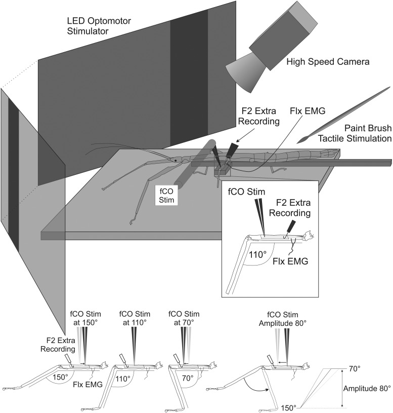
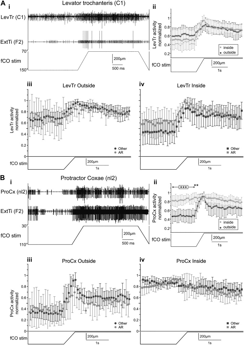
Feedback from load and movement sensors can modify timing and magnitude of the motor output in the stepping stick insect. One source of feedback is stretch reception by the femoral chordotonal organ (fCO), which encodes such parameters as the femorotibial (FTi) joint angle, the angular velocity, and its acceleration. Stimulation of the fCO causes a postural resistance reflex, during quiescence, and can elicit the opposite, so-called active reaction (AR), which assists ongoing flexion during active movements. In the present study, we investigated the role of fCO feedback for the difference in likelihood of generating ARs on the inside vs. the outside during curve stepping. We analyzed the effects of fCO stimulation on the motor output to the FTi and the neighboring coxa-trochanter and thorax-coxa joints of the middle leg. In inside and outside turns, the probability for ARs increases with increasing starting angle and decreasing stimulus velocity; furthermore, it is independent of the total angular excursion. However, the transition between stance and swing motor activity always occurs after a specific angular excursion, independent of the turning direction. Feedback from the fCO also has an excitatory influence on levator trochanteris motoneurons (MNs) during inside and outside turns, whereas the same feedback affects protractor coxae MNs only during outside steps. Our results suggest joint- and body side-dependent processing of fCO feedback. A shift in gain may be responsible for different AR probabilities between inside and outside turning, whereas the general control mechanism for ARs is unchanged.NEW & NOTEWORTHY We show that parameters of movement feedback from the tibia in an insect during curve walking are processed in a body side-specific manner, and how. From our results it is highly conceivable that the difference in motor response to the feedback supports the body side-specific leg kinematics during turning. Future studies will need to determine the source for the inputs that determine the local changes in sensory-motor processing.

Keywords: electrophysiology; motor control; reflex; sensorimotor; stick insect.


Conflict of interest statement

No conflicts of interest, financial or otherwise, are declared by the authors.

Figures

Fig. 1.
Fig. 1.
Curve walking and femoral chordotonal organ (fCO) stimulation setup. The animal is walking on a slippery surface while glued to a holder. The middle leg fCO is clamped and stimulated while flexor tibiae (FlxTi) electromyogram (EMG) signals from the same leg and the F2 nerve, containing extensor tibiae (ExtTi) motoneuron axons, are recorded extracellularly. Different starting angles (150°, 110°, and 70°) are applied by pre-stretching or pre-relaxing the receptor apodeme; changes in amplitude are applied by changing the height of the stimulation ramp, and velocities are varied by changing the slope of the ramp. Curve walking is induced visually and through tactile stimulation and is monitored by video. Stim, stimulation.
Fig. 2.
Fig. 2.
Example extensor tibiae (ExtTi) nerve and flexor tibiae (FlxTi) muscle recordings during inside and outside leg stepping with femoral chordotonal organ (fCO) stimulation (stim). Recordings of the ExtTi nerve (F2) and FlxTi electromyogram (EMG) are shown during ongoing inside (A) or outside (B) walking sequences (enlarged in Ci and Di, respectively) or during single fCO stimulations (Cii and Ciii; Dii and Diii), when the recorded side of the animal served as “inside” (A, Ci–Ciii) or “outside” (B, Di–Diii). Recordings in Cii, Ciii, and Dii show stimulations when active reactions (AR) were generated, and recording in Diii shows an undefined reaction. In Dii, single slow extensor tibiae (SETi) action potentials were observed at the very beginning of the ramp during outside steps (asterisk). Shaded areas indicate an actively walking animal (light gray, inside leg; dark gray, outside leg); open areas represent a resting animal. Black arrows marks part I and white arrows part II of an AR. CI, common inhibitor; FETi, fast extensor tibiae; RR, resistance reflex.
Fig. 3.
Fig. 3.
Stimulus time histograms of the extensor tibiae (ExtTi) motoneuron activity during femoral chordotonal organ (fCO) elongation. Plot shows the average action potential rate (AP rate, in AP/s) during the time window before, during, and after the fCO stimulus (stim) in a resting animal (i), active reactions (AR) in an active animal during inside stepping (ii; light gray) and outside stepping (iii; dark gray), and undefined activity during inside (iv) and outside stepping (v) (n = no. of observations).
Fig. 4.
Fig. 4.
Transition between mesothoracic (meso) flexor tibiae (FlxTi) and extensor tibiae (ExtTi) motoneuron activation during active reaction (AR). A: intracellular (intra) recording of the slow extensor tibiae motoneuron (SETi) and extracellular recordings of ExtTi nerve and FlxTi muscle showing an AR during an inside walking sequence. Black arrow marks beginning of part I and white arrow the beginning of part II of the AR. Angular difference until first AP is marked by shaded box. B: box-whisker plots of the angle at which the first ExtTi action potential (AP) occurred after stimulus (stim) onset for AR; x-axis gives value of the starting angle for the stimulation. C: box-whisker plots showing the angular difference between the stimulus onset and the first ExtTi AP for AR; x-axis gives value of the starting angle for the stimulation. Outside turn angles: 150° (N = 4 animals, n = 11 observations), 110° (N = 5, n = 14), 70° (N = 3, n = 50; inside turns: 150° (N = 6, n = 35), 110° (N = 5, n = 34), 70° (N = 4, n = 24). Light gray, inside leg; dark gray, outside leg. onset and first ExtTi AP are marked by black and white arrows, respectively. Box shows 25–75% interquartile range and median, whiskers are lower and upper 10% quartiles, and circles are outliers. **P < 0.01; ***P < 0.001.
Fig. 5.
Fig. 5.
Transition angle for an angular change of an extended 100° and all tested amplitudes together (40°, 60°, 80°, and 100°) for starting angles of 150° and 110° during outside and inside steps (see text for explanation). A and B: angle of the first extensor tibiae (ExtTi) action potential (AP) after stimulation (stim) onset for active reaction (AR) with a starting angle of 150° (left 2 boxes) and 110° (right 2 boxes) for outside steps (A) and inside steps (B). Outside number of animals (N) and number of observations (n): 150° (all amplitudes: N = 8, n = 47; 100° extended: N = 4, n = 33), 110° (all amplitudes: N = 6, n = 41; 100° extended: N = 4, n = 33). Inside N and n: 150° (all amplitudes: N = 7, n = 119; 100° extended: N = 6, n = 63); 110° (all amplitudes: N = 7, n = 93; 100° extended: N = 6, n = 61). Light gray, inside leg; dark gray, outside leg. Box shows 25–75% interquartile range and median, whiskers are lower and upper 10% quartiles, and dots are outliers.
Fig. 6.
Fig. 6.
Occurrence of active reaction (AR) for different stimulus (stim) parameters. A: occurrence of AR for inside [i; 70° (N = 13 animals, n = 441 observations), 110° (N = 21, n = 851), 150° (N = 18, n = 919)] and outside [ii; 70° (N = 8, n = 242), 110° (N = 18, n = 627), 150° (N = 15, n = 529)] middle leg for different starting angles. B: occurrence of AR for inside [i; 150°/s (N = 17, n = 656), 300°/s (N = 19, n = 731), 750°/s (N = 21, n = 843)] and outside [ii; 150°/s (N = 16, n = 448), 300°/s (N = 14, n = 447), 750°/s (N = 16, n = 481)] middle leg for different stimulus velocities. C: occurrence of AR for inside [i; 40° (N = 13, n = 724), 60° (N = 13, n = 750), 80° (N = 10, n = 373), 100° (N = 10, n = 387)] and outside [ii; 40° (N = 8, n = 406), 60° (N = 9, n = 432), 80° (N = 9, n = 299), 100° (N = 5, n = 265)] middle leg for different stimulus amplitudes. Light gray, inside leg; dark gray, outside leg. Box shows 25–75% interquartile range and median, whiskers are lower and upper 1.5 times interquartile range, and dots are outliers. *P < 0.05; **P < 0.01; ***P < 0.001.
Fig. 7.
Fig. 7.
Comparison of the occurrence of active reaction (AR) between inside and outside leg. Regression of the probability of occurrence of AR (solid lines) for different starting angles (i), different stimulus velocities (ii), and different stimulus amplitudes (iii). Dashed lines, 95% confidence intervals; light gray lines and circles, inside leg; dark gray lines and squares, outside leg. *P < 0.05; **P < 0.01.
Fig. 8.
Fig. 8.
Comparison of thoraco-coxal (ThC) and coxa-trochanteral (CTr) motoneuron activity as a result of femoral chordotonal organ (fCO) stimulation between inside and outside leg. Ai: recording of the levator trochanteris (LevTr) nerve (C1) and the extensor tibiae (ExtTi) nerve (F2) during fCO stimulation (stim), when the recorded side of the animal served as inside leg. Aii: comparison of normalized LevTr nerve activity during elongation stimuli at the fCO in the inside (light gray squares) or outside (dark gray circles) leg (N = 4 animals; inside leg, n = 150 observations; outside leg, n = 148 observations). Aiii and Aiv: comparison of normalized LevTr nerve activity during elongation stimuli at the fCO in the outside (iii) or inside leg (iv), but separated for the occurrence of AR (light gray; number of ARs: outside leg, nAR = 19; inside leg, nAR = 32) or other reaction (dark gray; number of other reactions: outside leg, nother = 129; inside leg, nother = 99) in the femorotibial (FTi) joint. No significant difference in the response was detected. Bi: recording of the protractor coxae (ProCx) nerve (nl2) and the ExtTi nerve (F2) during fCO stimulation, when the recorded side of the animal served as outside leg. Bii: comparison of normalized ProCx nerve activity during elongation stimuli at the fCO in the inside (light gray squares) or outside (dark gray circles) leg (N = 10; inside leg, n = 337; outside leg, n = 339). Biii and Biv: comparison of normalized ProCx nerve activity during elongation stimuli at the fCO in the outside (iii; N = 5) or inside leg (iv; N = 5), but separated for the occurrence of AR (light gray; outside leg, nAR = 53; inside leg, nAR = 110) or other reaction (dark gray; outside leg, nother = 168; inside leg, nother = 99) in the FTi joint. No significant difference in the response was detected. Note that fast ExtTi activity in Ai is much smaller than usual due to the extended starting position of the stimulus.

Similar articles

Cited by

References

    1. Bässler U. Reversal of a reflex to a single motoneuron in the stick insect Carausius morosus. Biol Cybern 24: 47–49, 1976. doi:10.1007/BF00365594. - DOI - PubMed
    1. Bässler U. Neural Basis of Elementary Behavior in Stick Insects. Berlin: Springer, 1983.
    1. Bässler U. Afferent control of walking movements in the stick insect Cuniculina impigra. I. Decerebrated animals on a treadband. J Comp Physiol A 158: 345–349, 1986a. doi:10.1007/BF00603618. - DOI
    1. Bässler U. Afferent control of walking movements in the stick insect Cuniculina impigra. II. Reflex reversal and the release of the swing phase in the restrained foreleg. J Comp Physiol 158: 351–362, 1986b. doi:10.1007/BF00603619. - DOI
    1. Bässler U. Functional principles of pattern generation for walking movements of stick insect forelegs: the role of the femoral chordotonal organ afferences. J Exp Biol 136: 125–147, 1988.

Publication types