

Developing a Highly Specific Biomarker for Germ Cell Malignancies: Plasma miR371 Expression Across the Germ Cell Malignancy Spectrum
- PMID: 31553692
- PMCID: PMC7351323
- DOI: 10.1200/JCO.18.02057
Developing a Highly Specific Biomarker for Germ Cell Malignancies: Plasma miR371 Expression Across the Germ Cell Malignancy Spectrum
Abstract
Purpose: Our objective was to evaluate operating characteristics, particularly specificity and positive predictive value (PPV), by mapping plasma miR371 expression to actual clinical events in patients with a history of germ cell tumor.
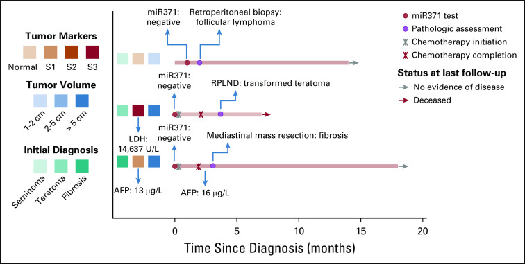
Patients and methods: One hundred eleven male patients with a history of or newly diagnosed germ cell tumors were evaluable. Biospecimens obtained before confirmed clinical events were analyzed for miR371 expression with blinding of providers and laboratory personnel to analytic results or clinical status, respectively. Cases (patients with clinically confirmed active germ cell malignancy [aGCM]) and controls (patients with no clinically confirmed aGCM) were assigned over the course of the management. Patients were assigned risk status (high, low, or moderate) based on the composite clinical picture at time points in management.
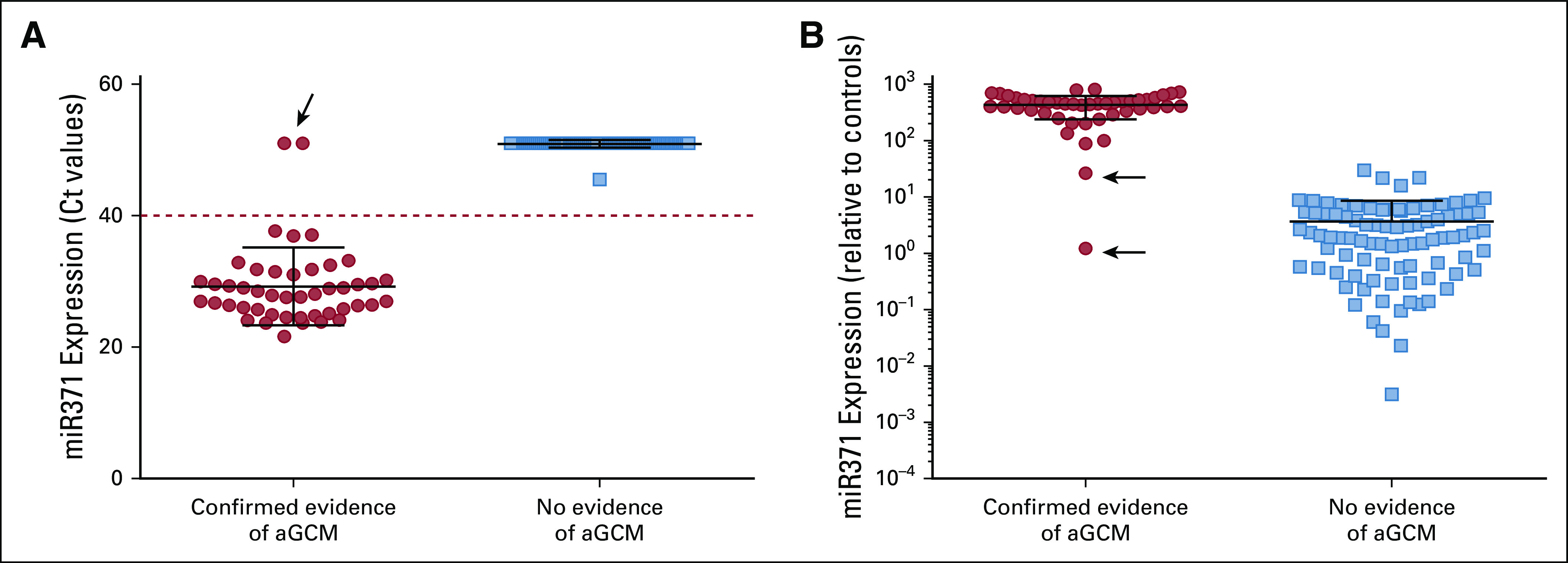
Results: Considering all cases and controls and results of prospectively obtained biosamples analyzed for miR371 expression, 46 (35%) of 132 samples had clinically confirmed aGCM over the course of management; 44 (96%) of these 46 patients had plasma miR371 expression (true positives) with no false positives. Two (4%) of 46 patients had no miRNA expression despite pathologic confirmation of aGCM (false negatives). Plasma miR371 expression in confirmed aGCM had a specificity, sensitivity, positive predictive value, and negative predictive value of 100%, 96%, 100%, and 98%, respectively. Interpretation of sensitivity and negative predictive value is limited by modest follow-up. Specificity and sensitivity were 100% and 98%, 100% and 92%, and 100% and 97% in the low-, moderate-, and high-risk groups, respectively, with a median follow-up time of 15 months.
Conclusion: Plasma miR371 expression predicts aGCM with high specificity and positive predictive value. Although other operating characteristics of miR371 await longer follow-up for more complete definition, the findings of a highly specific liquid biopsy strongly support moving forward with large-scale, real-world clinical trials to further define full operating characteristics and to identify clinical utility and areas of patient benefit.
Figures


Comment in
-
Refinement of the Management of Germ Cell Cancer: What's Next?J Clin Oncol. 2019 Nov 20;37(33):3063-3065. doi: 10.1200/JCO.19.02302. Epub 2019 Oct 4. J Clin Oncol. 2019. PMID: 31584840 No abstract available.
-
Re: Developing a Highly Specific Biomarker for Germ Cell Malignancies: Plasma miR371 Expression across the Germ Cell Malignancy Spectrum.J Urol. 2020 May;203(5):884. doi: 10.1097/JU.0000000000000780.01. Epub 2020 Feb 11. J Urol. 2020. PMID: 32073935 No abstract available.
-
Re: Developing a Highly Specific Biomarker for Germ Cell Malignancies: Plasma miR371 Expression across the Germ Cell Malignancy Spectrum.J Urol. 2020 May;203(5):884. doi: 10.1097/JU.000000000000078.01. Epub 2020 Feb 11. J Urol. 2020. PMID: 32264776 No abstract available.
References
-
- Einhorn LH. Treatment of testicular cancer: A new and improved model. J Clin Oncol. 1990;8:1777–1781. - PubMed
-
- Eini R, Dorssers LC, Looijenga LH. Role of stem cell proteins and microRNAs in embryogenesis and germ cell cancer. Int J Dev Biol. 2013;57:319–332. - PubMed
-
- Gillis AJ, Stoop HJ, Hersmus R, et al. High-throughput microRNAome analysis in human germ cell tumours. J Pathol. 2007;213:319–328. - PubMed
-
- Voorhoeve PM, le Sage C, Schrier M, et al. A genetic screen implicates miRNA-372 and miRNA-373 as oncogenes in testicular germ cell tumors. Cell. 2006;124:1169–1181. - PubMed
Publication types
MeSH terms
Substances
Grants and funding
LinkOut - more resources
Full Text Sources
Other Literature Sources
