. 2019 Sep 25;14(9):e0222924.
doi: 10.1371/journal.pone.0222924. eCollection 2019.

NPHP proteins are binding partners of nucleoporins at the base of the primary cilium

Affiliations

NPHP proteins are binding partners of nucleoporins at the base of the primary cilium

T Lynne Blasius et al. PLoS One. .

Abstract

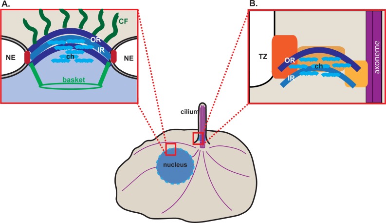
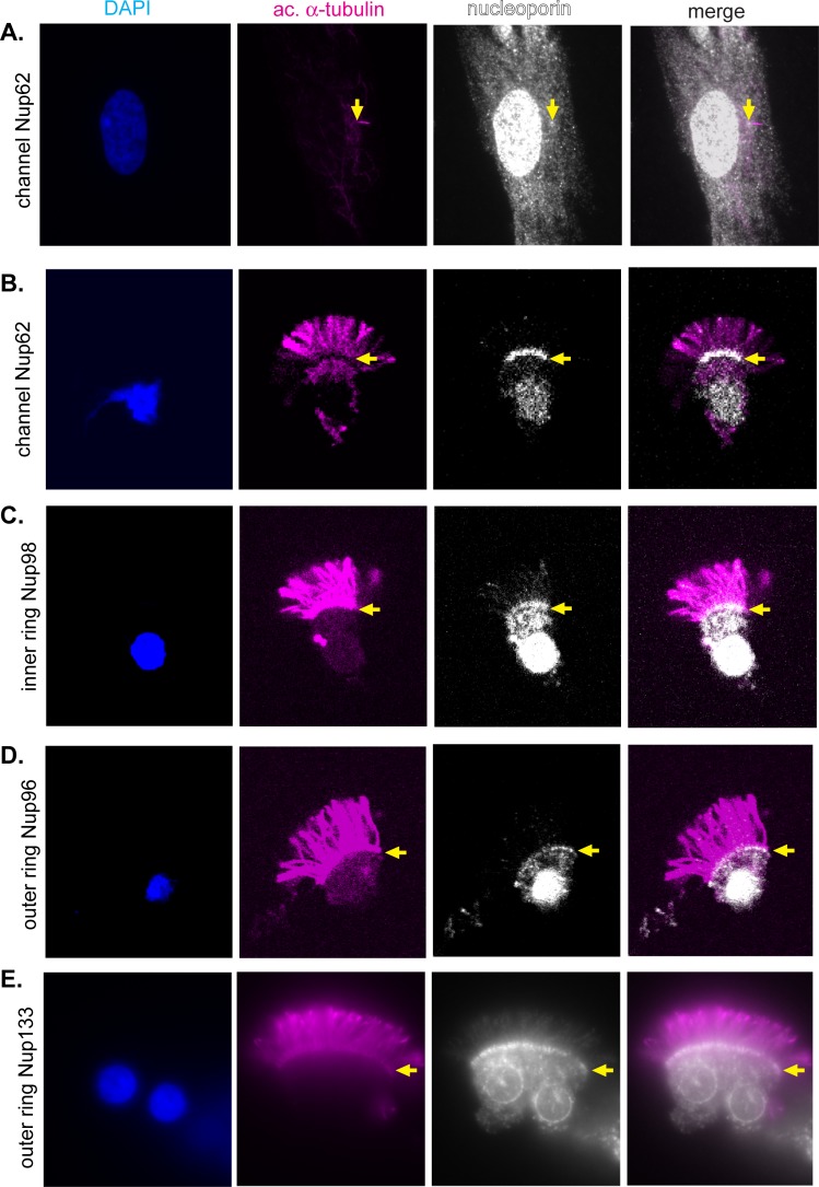
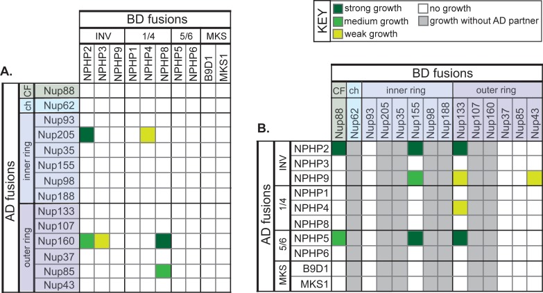
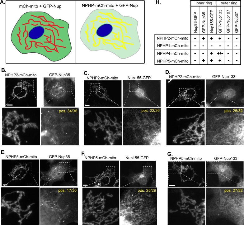
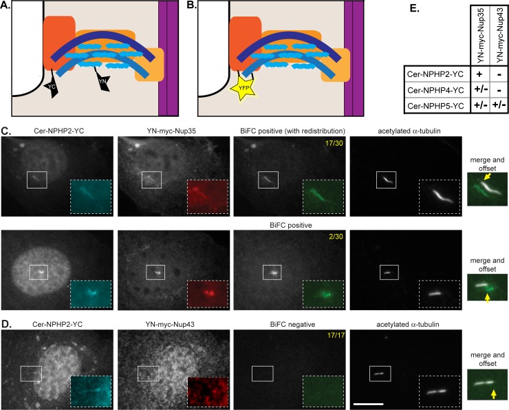
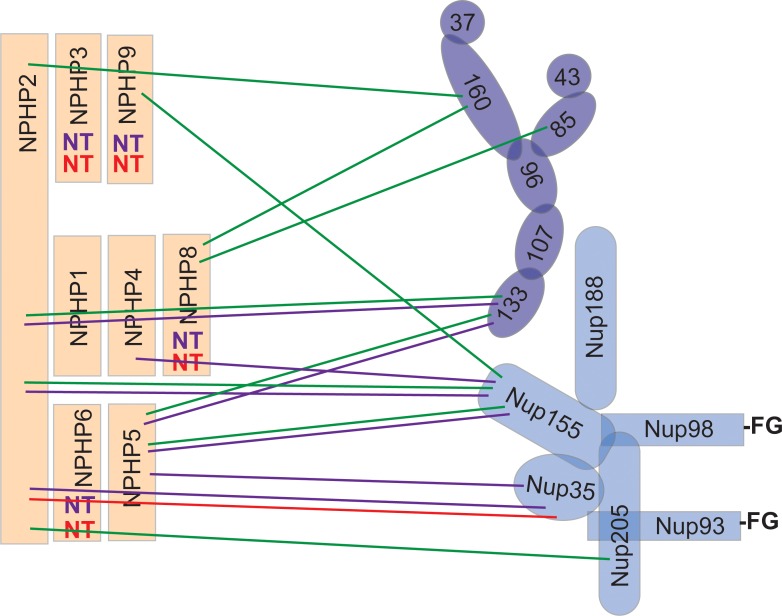
Cilia are microtubule-based organelles that protrude from the surface of eukaryotic cells to generate motility and to sense and respond to environmental cues. In order to carry out these functions, the complement of proteins in the cilium must be specific for the organelle. Regulation of protein entry into primary cilia has been shown to utilize mechanisms and components of nuclear gating, including nucleoporins of the nuclear pore complex (NPC). We show that nucleoporins also localize to the base of motile cilia on the surface of trachea epithelial cells. How nucleoporins are anchored at the cilium base has been unclear as transmembrane nucleoporins, which anchor nucleoporins at the nuclear envelope, have not been found to localize at the cilium. Here we use the directed yeast two-hybrid assay to identify direct interactions between nucleoporins and nephronophthisis proteins (NPHPs) which localize to the cilium base and contribute to cilium assembly and identity. We validate NPHP-nucleoporin interactions in mammalian cells using the knocksideways assay and demonstrate that the interactions occur at the base of the primary cilium using bimolecular fluorescence complementation. We propose that NPHP proteins anchor nucleoporins at the base of primary cilia to regulate protein entry into the organelle.


Conflict of interest statement

The authors have declared that no competing interests exist.

Figures

Fig 1
Fig 1. Schematic of nuclear and ciliary gating complexes.
(A) At the nuclear envelope (NE), nucleoporins in NPCs are organized into subcomplexes including outer ring (OR, dark blue), inner ring (IR, medium blue), central channel (ch, light blue), cytoplasmic filaments (CF, dark green), nuclear basket (green), and transmembrane (red). (B) At the base of the primary cilium, OR, IR and channel nucleoporins localize at the transition zone (TZ) with NPHP proteins (orange). Purple lines indicate microtubules.
Fig 2
Fig 2. Inner ring, outer ring, and central channel nucleoporins localize to the base of motile cilia.
(A) RPE-1 cells were stained with antibodies to the channel nucleoporin Nup62 (gray). (B-E) Epithelial cells isolated from rat trachea were stained with antibodies against (b) the channel nucleoporin Nup62, (C) the inner ring nucleoporin Nup98, or the outer ring nucleoporins (D) Nup96 and (E) Nup133. All cells were counter stained with antibodies to acetylated α-tubulin (magenta) to mark the ciliary axoneme and with DAPI (blue) to mark nuclei. Yellow arrows indicate the base of the cilia.
Fig 3
Fig 3. Directed yeast two-hybrid assay reveals NUP-NPHP interactions.
The indicated transition zone proteins (INV, NPHP1/4, NPHP5/6, MKS complexes) and nucleoporin proteins were fused to a DNA binding domain (BD) or transcription activation domain (AD). Yeast of different mating types were mated pairwise to test for protein-protein interactions. Transcriptional activation allowing growth of diploid yeast was observed after 3 days and scored as strong growth (dark green), medium growth (medium green), or weak growth (yellow-green). Each pairwise mating was repeated at least three times. CF, cytoplasmic filament nucleoporin. ch, central channel nucleoporin.
Fig 4
Fig 4. Identification of NPHP-NUP interactions by the knocksideways assay.
(A) Schematic of the knocksideways assay. NPHP proteins were tagged with mCherry (mCh) and targeted to the mitochondrial surface by fusion to a mitochondrial targeting sequence (mito). The NPHP-mCh-mito bait was co-expressed with an EGFP-tagged nucleoporin (GFP-Nup). An interaction between the NPHP and nucleoporin proteins recruits the GFP-Nup to the mCh-labeled mitochondrial surface. (B-D) NPHP2-mCh-mito recruits the inner ring nucleoporins (B) GFP-Nup35 and (C) Nup155-EGFP3 and the outer ring nucleoporin (D) GFP-Nup133 to the mitochondrial surface. (E-G) NPHP5-mCh-mito recruits the inner ring nucleoporins (E) GFP-Nup35 and (F) Nup155-EGFP3 and the outer ring nucleoporin (G) GFP-Nup133 to the mitochondrial surface. Scale bars, 10 μm. Yellow text indicates the number of cells positive (pos.) for a knocksideways interaction. (H) Summary of the results. +, recruitment of GFP-Nup observed in >50% of cells; +/-, recruitment of GFP-Nup observed in 10–50% of cells; -, recruitment of GFP-Nup observed in <10% of cells.
Fig 5
Fig 5. BiFC analysis reveals close association of NPHP2 and Nup35 at the base of the cilium.
(A-B) Schematic of the assay. NPHP proteins (orange) were tagged with a C-terminal fragment of YFP (YC) and with Cerulean (Cer) to detect protein expression. Nucleoporin proteins (blue) were tagged with an N-terminal fragment of YFP (YN) and with a Myc-tag to detect protein expression. (A) When co-expressed in NIH 3T3 cells, the NPHP-YC and YN-NUP proteins localize to the base of the cilium but if they are not in close proximity, the YFP protein is not reconstituted (no BiFC). (B) If the NPHP-YC and YN-NUP proteins are in close proximity at the base of the cilium, then YFP is reconstituted and BiFC fluorescence is observed (right panel). (C) Co-expression of Cer-NPHP2-YC with the inner ring nucleoporin YN-myc-Nup35 results in a positive BiFC interaction. (D) Co-expression of Cer-NPHP2-YC with the outer ring nucleoporin YN-myc-Nup43 results in a positive BiFC interaction. Scale bar, 5 um. Yellow text in the top right corners indicate the number of cells displaying positive BiFC interactions across two independent experiments. Yellow arrows indicate the base of the primary cilium. (E) Summary of BiFC interactions observed for the indicated NPHP and nucleoporin proteins.
Fig 6
Fig 6. Summary of NPHP-NUP interactions.
Components of the inner ring nucleoporin (medium blue), outer ring nucleoporin (dark blue), and NPHP (orange) complexes are indicated. Green lines indicate interactions detected in the directed yeast two-hybrid assay (strong and medium only). Purple lines indicate interactions detected in the knocksideways assays. The red line indicates the spatial proximity relationship observed in the BiFC assay. FG, Phenylalanine-Glycine repeats. NT, not tested.

References

    1. Anvarian Z, Mykytyn K, Mukhopadhyay S, Pedersen LB, Christensen ST (2019) Cellular signalling by primary cilia in development, organ function and disease. Nat Rev Nephrol 15: 199–219. 10.1038/s41581-019-0116-9 - DOI - PMC - PubMed
    1. Elliott KH, Brugmann SA (2019) Sending mixed signals: Cilia-dependent signaling during development and disease. Dev Biol 447: 28–41. 10.1016/j.ydbio.2018.03.007 - DOI - PMC - PubMed
    1. Mitchison HM, Valente EM (2017) Motile and non-motile cilia in human pathology: from function to phenotypes. J Pathol 241: 294–309. 10.1002/path.4843 - DOI - PubMed
    1. Braun DA, Hildebrandt F (2017) Ciliopathies. Cold Spring Harb Perspect Biol 9:a028191 10.1101/cshperspect.a028191 - DOI - PMC - PubMed
    1. Reiter JF, Leroux MR (2017) Genes and molecular pathways underpinning ciliopathies. Nat Rev Mol Cell Biol 18: 533–547. 10.1038/nrm.2017.60 - DOI - PMC - PubMed

Publication types

MeSH terms

Substances