. 2019 Oct 24;62(20):9201-9216.
doi: 10.1021/acs.jmedchem.9b01120. Epub 2019 Oct 11.

Anti-biofilm Agents against Pseudomonas aeruginosa: A Structure-Activity Relationship Study of C-Glycosidic LecB Inhibitors

Affiliations

Anti-biofilm Agents against Pseudomonas aeruginosa: A Structure-Activity Relationship Study of C-Glycosidic LecB Inhibitors

Roman Sommer et al. J Med Chem. .

Abstract

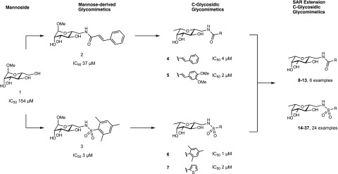
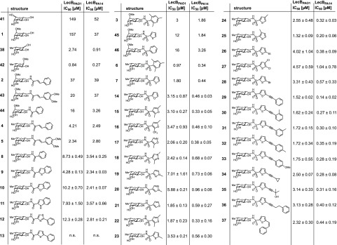
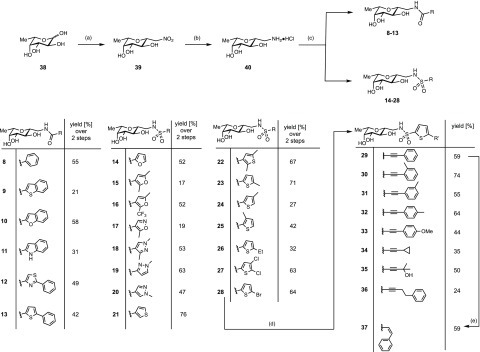
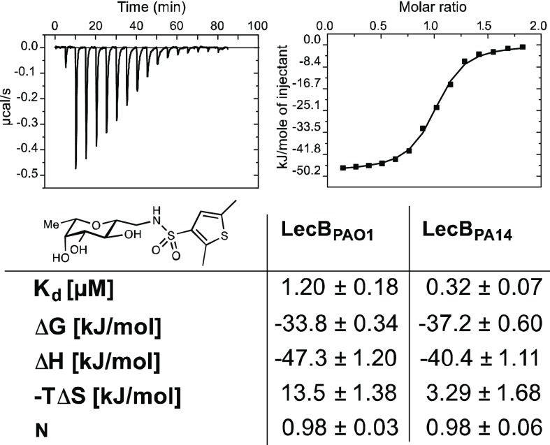
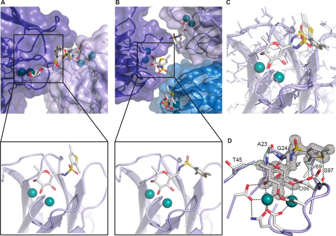
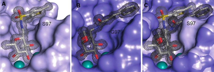
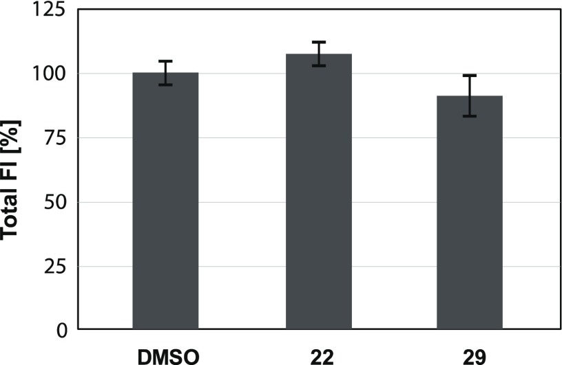
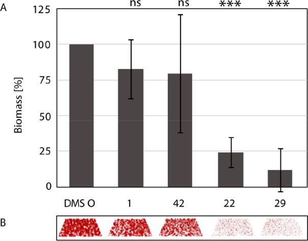
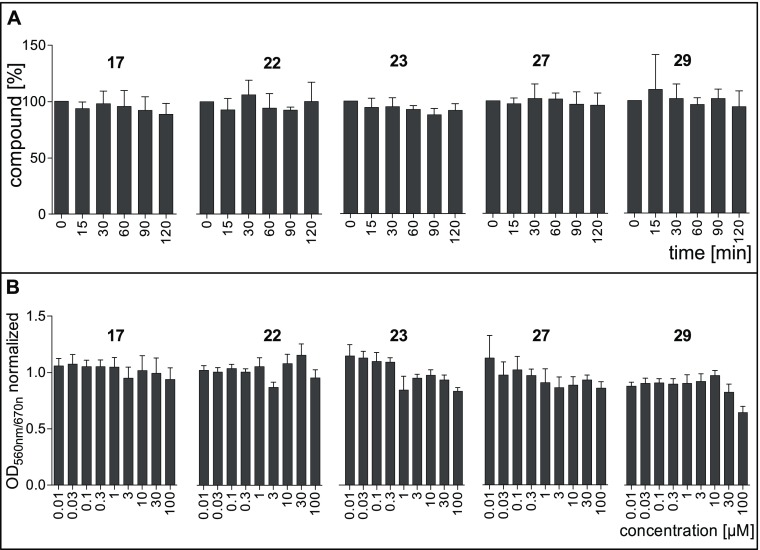
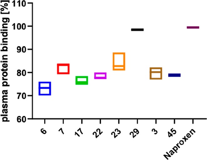
Biofilm formation is a key mechanism of antimicrobial resistance. We have recently reported two classes of orally bioavailable C-glycosidic inhibitors of the Pseudomonas aeruginosa lectin LecB with antibiofilm activity. They proved efficient in target binding, were metabolically stable, nontoxic, selective, and potent in inhibiting formation of bacterial biofilm. Here, we designed and synthesized six new carboxamides and 24 new sulfonamides for a detailed structure-activity relationship for two clinically representative LecB variants. Sulfonamides generally showed higher inhibition compared to carboxamides, which was rationalized based on crystal structure analyses. Substitutions at the thiophenesulfonamide increased binding through extensive contacts with a lipophilic protein patch. These metabolically stable compounds showed a further increase in potency toward the target and in biofilm inhibition assays. In general, we established the structure-activity relationship for these promising antibiofilm agents and showed that modification of the sulfonamide residue bears future optimization potential.


Conflict of interest statement

The authors declare no competing financial interest.

Figures

Figure 1
Figure 1
Design approach for C-glycosidic LecB inhibitors and extension of the structural space for extended SAR studies of compounds 437. Derivatives of methyl α-d-mannoside 13 and their inhibitory potency for the binding with LecBPAO1.C-Glycosides simultaneously derived of d-mannosides and l-fucosides are hybrid-type LecB ligands 4 and 5 and 6 and 7.
Figure 2
Figure 2
Competitive binding assay of inhibitors with LecBPAO1 and LecBPA14 based on fluorescence polarization. Means and standard deviations were determined from a minimum of three independent experiments. n.s.: not soluble at 1 mM in TBS/Ca containing 1% DMSO. IC50 values for 17, 38, and 4146 with LecBPAO1 and LecBPA14 were previously published.,−,
Scheme 1
Scheme 1. Synthesis of the Amides 813 and Sulfonamides 1438
Reagents and conditions: (a) MeNO2, DBU, molecular sieves 3 Å, 1,4-dioxane, 50 °C, 3 d; (b)Pt/C, H2, HCl, MeOH, rt, 2 d; (c) acyl/sulfonyl chloride or carboxylic acid/EDC·HCl, Et3N, DMF, 0 °C; (d) CuI, Pd(PPh3)2Cl2, RCCH, Et3N, DMF, 50 °C, 16–42 h; (e) 1 atm H2, Lindlar’s catalyst, quinoline, rt, 46 h. Yields for 828 are given over two steps from the nitro derivative 39.
Figure 3
Figure 3
Isothermal titration microcalorimetry of LecBPAO1 and LecBPA14 with dimethylthiophene 22. Means and standard deviations were determined from a minimum of three independent titrations. One representative titration graph is depicted for LecBPAO1 only.
Figure 4
Figure 4
Crystal structure of LecBPA14 with C-glycoside ligand 22 (1.45 Å resolution, PDB 5MAZ), (A) Observed binding pose of 22 with crystal contacts. (B) Observed binding pose of 22 without crystal contacts. (C) Superposition of both observed binding poses. (D) Relevant binding pose of 22 with 2FobsFcalc electron density displayed at 1σ. Ligands and amino acids of the carbohydrate recognition domain (CRD) are depicted as sticks colored by elements (C, gray; N, blue; O, red; S, yellow); protein surface in transparent blue and two Ca2+-ions in the binding sites are shown as green spheres.
Figure 5
Figure 5
(A) Crystal structure of the complex of LecBPA14 with dimethylthiophene 22 (PDB 5MAZ) reveals the hydrophobic interaction of the ortho-methyl group attached to the thiophene residue with a hydrophobic patch on the protein surface. Furthermore, the thiophene interacts with the side chain CH2 of Ser97. (B) Crystal structure of the complex of LecBPAO1 with manno-cinnamide 2 (PDB 5A3O) reveals the hydrophobic interaction of the cinnamoyl group with a hydrophobic patch formed by the loop carrying Gly97 that lacks the serine side chain present in LecBPA14. (C) superposition of the two structures indicating the steric clash between carboxamide substituents, conformationally fixed through a hydrogen bond of their NH group with the carboxylate of Asp96, and the bulk of the side chain of Ser97 in LecBPA14. This serves as an explanation for the increased selectivity of the amides for LecBPAO1, which contrasts the selectivity of the sulfonamides for LecBPA14. Electron density 2FobsFcalc is displayed at 1σ. Ligands are depicted as sticks colored by elements (C, gray; N, blue; O, red; S, yellow); protein surface in blue and two Ca2+-ions in the binding sites are shown as green spheres.
Figure 6
Figure 6
Bacterial growth of mCherry-expressing P. aeruginosa quantified by fluorescence intensity (FI) and normalized on the DMSO control in the presence of 100 μM lectin inhibitors 22 or 29.
Figure 7
Figure 7
Inhibition of biofilm formation by P. aeruginosa after 48 h growth in the presence of compounds 22 or 29. Depicted data for methyl α-l-fucoside (42), 1, and the DMSO control have been published. (A) Quantification of biofilm biomass. Averages and standard deviations of biofilm formation from three independent assays. Statistical significance was calculated using the Student’s t test. (B) Raw data of confocal fluorescence microscopy 3D images show one representative z-stack per condition.
Figure 8
Figure 8
(A) Stability of LecB ligands in mouse plasma. The error bars show the standard deviation of minimum three assays. (B) Toxicity of LecB ligands to human liver Hep G2 cells. Measured OD560nm/670nm was normalized to the controls. Untreated cells served as negative control (normalized OD560nm/670nm = 1), and Triton X-100 treated cells served as positive control (normalized OD560nm/670nm = 0). The error bars show the standard deviation of minimum three assays.
Figure 9
Figure 9
Plasma protein binding of LecB inhibitors C-glycosides 629 and the O-glycosides 3 and 45 and naproxen as a control. Box plots show mean and confidence intervals.

References

    1. Nagao M.; Iinuma Y.; Igawa J.; Saito T.; Yamashita K.; Kondo T.; Matsushima A.; Takakura S.; Takaori-Kondo A.; Ichiyama S. Control of an outbreak of carbapenem-resistant Pseudomonas aeruginosa in a haemato-oncology unit. J. Hosp Infect 2011, 79, 49–53. 10.1016/j.jhin.2011.04.018. - DOI - PubMed
    1. Rice L. B. Federal funding for the study of antimicrobial resistance in nosocomial pathogens: No ESKAPE. J. Infect. Dis. 2008, 197, 1079–1081. 10.1086/533452. - DOI - PubMed
    1. Tsutsui A.; Suzuki S.; Yamane K.; Matsui M.; Konda T.; Marui E.; Takahashi K.; Arakawa Y. Genotypes and infection sites in an outbreak of multidrug-resistant Pseudomonas aeruginosa. J. Hosp Infect 2011, 78, 317–322. 10.1016/j.jhin.2011.04.013. - DOI - PubMed
    1. WHO Publishes List of Bacteria for Which New Antibiotics Are Urgently Needed; World Health Organization: Geneva, 2017; http://www.who.int/mediacentre/news/releases/2017/bacteria-antibiotics-n... (accessed May 2017)
    1. Hauser A. R., Rello J., Eds. Severe Infections Caused by Pseudomonas aeruginosa; Kluwer Academic Publishers Group: Boston, MA, 2003.

Publication types

MeSH terms