. 2019 Sep 24;28(13):3462-3473.e5.
doi: 10.1016/j.celrep.2019.08.058.

hESC-Derived Dopaminergic Transplants Integrate into Basal Ganglia Circuitry in a Preclinical Model of Parkinson's Disease

Affiliations

hESC-Derived Dopaminergic Transplants Integrate into Basal Ganglia Circuitry in a Preclinical Model of Parkinson's Disease

Andrew F Adler et al. Cell Rep. .

Abstract

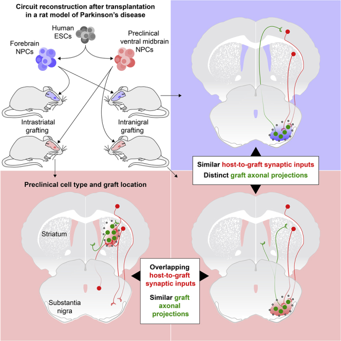
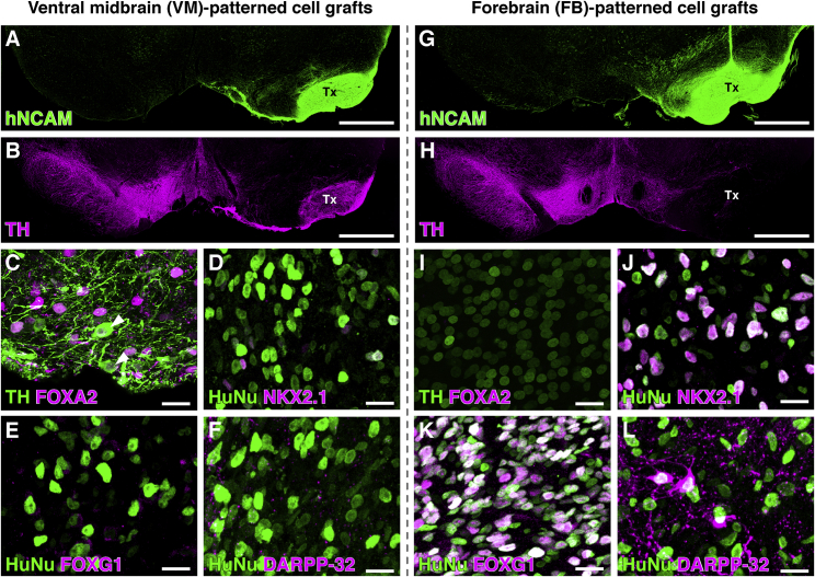
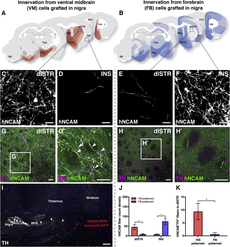
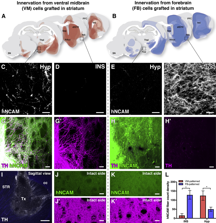
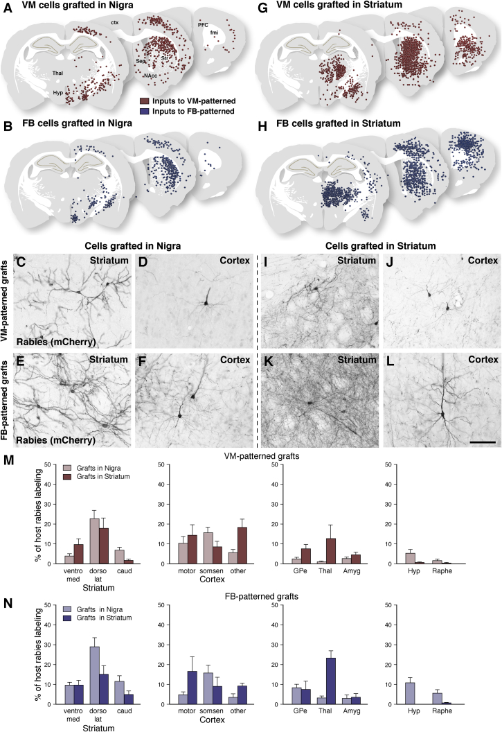
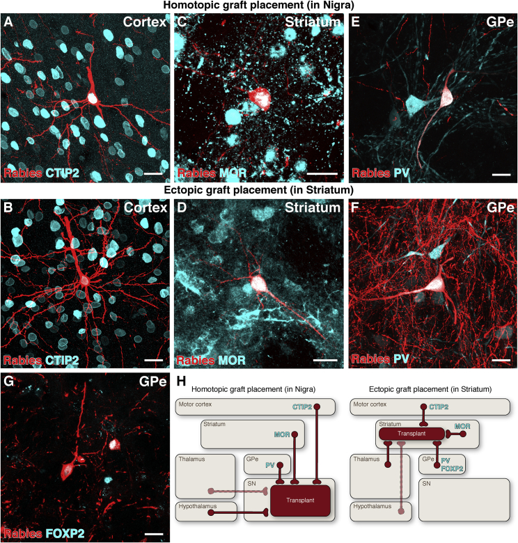
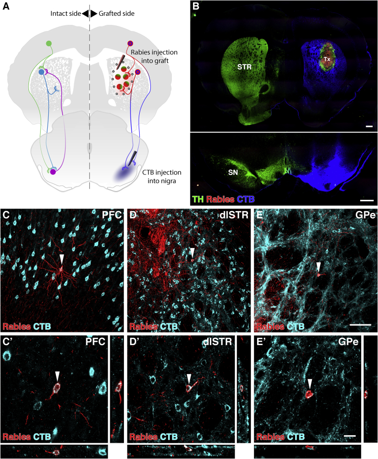
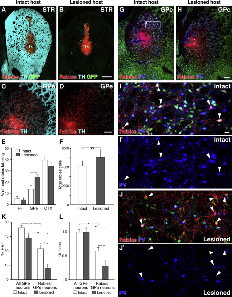
Cell replacement is currently being explored as a therapeutic approach for neurodegenerative disease. Using stem cells as a source, transplantable progenitors can now be generated under conditions compliant with clinical application in patients. In this study, we elucidate factors controlling target-appropriate innervation and circuitry integration of human embryonic stem cell (hESC)-derived grafts after transplantation to the adult brain. We show that cell-intrinsic factors determine graft-derived axonal innervation, whereas synaptic inputs from host neurons primarily reflect the graft location. Furthermore, we provide evidence that hESC-derived dopaminergic grafts transplanted in a long-term preclinical rat model of Parkinson's disease (PD) receive synaptic input from subtypes of host cortical, striatal, and pallidal neurons that are known to regulate the function of endogenous nigral dopamine neurons. This refined understanding of how graft neurons integrate with host circuitry will be important for the design of clinical stem-cell-based replacement therapies for PD, as well as for other neurodegenerative diseases.

Keywords: Parkinson’s disease; brain repair; cell replacement therapy; cell transplantation; grafting; monosynaptic tracing; stem cells.


Conflict of interest statement

M.P. is the owner of Parmar Cells AB and co-inventor of the U.S. patent application 15/093,927 owned by Biolamina AB and EP17181588 owned by Miltenyi Biotech.

Figures

None
Graphical abstract
Figure 1
Figure 1
Comparison of VM- versus FB-Patterned Graft Composition Phenotypic characterization of hESC-derived VM-patterned (A–F) and FB-patterned cells (G–L) 6 months after grafting to the nigra. (A, B, G, and H) VM- (A) and FB-patterned cells (G) generated grafts of comparable size, as visualized by human NCAM staining. VM- (B) but not FB-patterned grafts (H) generated TH+ neurons in vivo. (C and I) In VM-patterned grafts (C) but not FB-patterned grafts (I), TH+ neurons also co-expressed the midbrain DA neuron marker FOXA2 (arrowheads). (D–F and J–L) FB- (J) but not VM-patterned graft cells (HuNu+) (D) expressed NKX2.1 and FOXG1 (E and K), and included DARPP-32+ neurons (F and L). See also Figure S1 for in vitro characterization of cell preparations. All graft neurons expressed a rabies tracing construct and, thus, nuclear GFP. Scale bars represent 1 mm (A, B, G, and H) and 20 μm (C–F and I–L). Images in (A), (B), (G), and (H) are digitally stitched from multiple high-magnification images. DARPP-32, dopamine- and cAMP-regulated phosphoprotein; FB, forebrain; FOXA2, forkhead box A2; FOXG1, forkhead box protein G1; hESCs, human embryonic stem cells; HuNu, human nucleus; NKX2.1, NK2 homeobox 1; TH, tyrosine hydroxylase; Tx, transplant; VM, ventral midbrain.
Figure 2
Figure 2
Comparison of Fiber Outgrowth from VM- versus FB-Patterned Grafts Placed into the Substantia Nigra (A and B) Figures showing composite data of graft-derived innervation of host structures as detected by DAB-developed hNCAM staining for grafts placed in the nigra of 6-OHDA lesioned rats after 24 weeks of graft maturation (combines and visualizes data collected from n = 5 animals in A and n = 6 animals in B). Data from the group of animals in (A) have also been used in Cardoso et al., 2018. (C–F) VM-patterned neurons targeted fiber outgrowth toward dorsolateral striatum (dlSTR) (C) but not insular cortex (INS) (D), while FB-patterned neurons extensively INS (F) but not dlSTR (E). (G) Human (hNCAM+) TH+ fibers reinnervated the dopamine-depleted striatum from VM-patterned grafts. (H) Comparatively sparse TH fibers extended into the striatum from FB-patterned grafts. (I) Tissue clearing and light sheet microscopy provides an overview of graft-derived TH+ fibers extending to reinnervate the DA-depleted host striatum after 16 weeks of maturation, with endogenous autofluorescence shown in blue (see associated Videos S1 and S2). (J and K) Quantitative image analysis of hNCAM+ (J) and hNCAM+TH+ (K) fiber outgrowth confirms that 24-week VM-patterned grafts preferentially innervated the dlSTR, whereas FB-patterned grafts preferentially innervated insular cortex (VM cells, n = 5 animals; FB cells, n = 6 animals; mean ± SEM, p < 0.05, α = 0.05, two-way ANOVA with Bonferroni-corrected post hoc testing for hNCAM, unpaired two-tailed t test for hNCAM+TH+). Scale bars represent 20 μm (C–F), 50 μm (G and H), and 500 μm (I, sagittal plane). 6-OHDA, 6-hydroxydopamine; DAB, 3,3′-diaminobenzidine; dlSTR, dorsolateral striatum; fmi, forceps minor; FB, forebrain; hNCAM, human neural cell adhesion molecule; Hyp, hypothalamus; INS, insular cortex; MFB, medial forebrain bundle; NAcc, nucleus accumbens; PFC, prefrontal cortex; Sep, septum; SN, substantia nigra; TH, tyrosine hydroxylase; Thal, thalamus; Tx, transplant; VM, ventral midbrain.
Figure 3
Figure 3
Comparison of Fiber Outgrowth from VM- versus FB-Patterned Grafts Placed into the Striatum (A and B) Representations of graft-derived innervation of host structures as detected by hNCAM staining for grafts placed in the striatum of 6-OHDA lesioned rats after 24 weeks of graft maturation (cumulative data from n = 7 animals in A and n = 5 animals in B). (C–F) VM-patterned neurons targeted fiber outgrowth toward the hypothalamus (Hyp) (C) but not INS (D), while FB-patterned neurons extensively INS (F) but not Hyp (E). (G and H) Human (hNCAM+) TH+ fibers reinnervated the dopamine-depleted striatum from VM-patterned grafts (G), but not from FB-patterned grafts (H), after 24 weeks of maturation. (I) Tissue clearing and light sheet microscopy provide an overview of VM graft-derived TH+ fibers reinnervating the DA-depleted host striatum after 16 weeks of maturation, with endogenous autofluorescence shown in blue (see associated Video S3). (J and K) Images of the contralateral (intact) side of the striatum in animals with (J) VM- and (K) FB-patterned grafts confirm the specificity of hNCAM labeling. (L) Quantification of VM- versus FB-patterned hNCAM+ innervation of host structures. (VM cells, n = 7 animals; FB cells, n = 5 animals; mean ± SEM, p < 0.05, α = 0.05, two-way ANOVA with Bonferroni-corrected post hoc testing). Scale bars represent 20 μm (C–F), 50 μm (G, H, J, and K), and 500 μm (I, sagittal plane). The TH channel in (G) and (H) are displayed with a higher exposure time than (J) and (K) to prevent over- and undersaturation. 6-OHDA, 6-hydroxydopamine; cc, corpus callosum; dlSTR, dorsolateral striatum; fmi, forceps minor; FB, forebrain; hNCAM, human neural cell adhesion molecule; Hyp, hypothalamus; INS, insular cortex; MFB, medial forebrain bundle; NAcc, nucleus accumbens; PFC, prefrontal cortex; Sep, septum; SN, substantia nigra; STR, striatum; TH, tyrosine hydroxylase; Thal, thalamus; Tx, transplant; VM, ventral midbrain.
Figure 4
Figure 4
Role of Graft Phenotype and Placement on Synaptic Integration (A–L) Schematic overviews (A, B, G, and H) of (C)–(F) and (I)–(L) 3,3′-diaminobenzidine (DAB)-developed staining of host synaptic inputs to VM- and FB-patterned grafts placed in either the nigra (A–F) or striatum (G–L). Each dot represents a ΔG-rabies+ traced neuron tagged with mCherry collected from 1:8 series spanning 2.0, 1.4, or 2.4 mm (rostral to caudal) centered at each depicted section, cumulative from all animals in each group. Synaptic inputs to VM- and FB-patterned neurons originated from the same host structures when placed in the same location (compare A to B and G to H). On the other hand, synaptic inputs varied depending on the placement of the transplant (compare A to G and B to H). (A) Inputs to VM-patterned grafts placed in the nigra included host (C) striatal and (D) cortical neurons. (B) Inputs to FB-patterned grafts placed in the nigra included host (E) striatal and (F) cortical neurons. (G) Inputs to VM-patterned grafts placed in the striatum included host (I) striatal and (J) cortical neurons. (H) Inputs to FB-patterned grafts placed in the striatum included host (K) striatal and (L) cortical neurons. (M and N) Quantification of the percentage of rabies+ neurons per anatomical structure over the total number of ΔG-rabies+ neurons per brain in animals grafted with (M) VM- or (N) FB-patterned cells (VM cells in SN, n = 5 animals; FB cells in SN, n = 6 animals; VM cells in striatum, n = 7 animals; FB cells in striatum, n = 5 animals). Data presented as mean + SEM. See also Figure S2. Scale bar represents 20 μm (C–F and I–L). Amyg, amygdala; caud, caudal striatum; ctx, cortex; dorso lat, dorsolateral striatum; FB, forebrain; fmi, forceps minor; GPe, external globus pallidus; Hyp, hypothalamus; motor, motor cortex; NAcc, nucleus accumbens; PFC, prefrontal cortex; sep, septum; somsen, somatosensory cortex; Str, striatum; Thal, thalamus; VM, ventral midbrain; ventro med, ventromedial striatum.
Figure 5
Figure 5
Phenotypic Similarity of Monosynaptic Inputs to Ectopic versus Homotopic VM-Patterned Grafts (A–F) CTIP2+ pyramidal neurons in motor cortex (A and B), MOR+ medium spiny neurons in striatum (C and D), and PV+ neurons in GPe (E, F) connected to both homotopic (intranigral) and ectopic (intrastriatal) grafts of VM-patterned neurons. (G) FOXP2+ GPe neurons were observed connecting only to grafts placed ectopically in the striatum. (H) Schematic representations of the origin of host synaptic inputs to grafts placed in the substantia nigra versus the striatum. Solid lines represent host brain structures with extensive synaptic input to transplanted neurons, while dashed lines represent structures with comparatively scarce inputs. Scale bars represent 20 μm. CTIP2, COUP-TF-interacting protein 2; FOXP2, forkhead box P2; GPe, external globus pallidus; MOR, mu-opioid receptor; PV, parvalbumin; SN, substantia nigra.
Figure 6
Figure 6
Host Neurons Providing Monosynaptic Input to VM-Patterned Grafts Placed in the Striatum Simultaneously Collateralize on Neurons in the Substantia Nigra (A) Schematic of monosynaptic EnvA ΔG-rabies tracing initiated from striatal grafts, with simultaneous conventional retrograde cholera toxin subunit B (CTB) tracing of projections to the substantia nigra. (B) Sections through striatum depicting the graft and rabies injection site, and the substantia nigra depicting the lesion and CTB injection site. (C–E) Host prefrontal cortical (C), medium spiny striatal (D), and pallidal neurons (E) were labeled with both CTB and rabies. Arrowheads indicate neurons imaged with confocal microscopy in (C’), (D’), and (E’). These neurons simultaneously synapse with graft neurons and maintain a collateral projection to the substantia nigra. Scale bars represent 500 μm (B), 100 μm (C, D, and E), and 20 μm (C’, D’, and E’). Images in (B) is digitally stitched from multiple high-magnification images. CTB, cholera toxin subunit B; dlSTR, dorsolateral striatum; GPe, external globus pallidus; PFC, prefrontal cortex; SN, substantia nigra; STR, striatum; TH, tyrosine hydroxylase; Tx, transplant.
Figure 7
Figure 7
Monosynaptic Inputs to VM-Patterned Grafts in Intact versus 6-OHDA Lesioned Rats (A and B) VM-patterned cells placed in the striatum of intact (A) or 6-OHDA-lesioned rats (B) generate grafts of comparable size. (C–E) Lesioned host animals had an increased number of rabies-labeled neurons in the GPe (C and D), as well as an increased fraction of GPe inputs normalized to labeling in the entire brain (E). (F) The overall number of ΔG-rabies-labeled host neurons was not significantly different between intact and lesioned hosts. (G–J) ΔG-rabies-labeled GPe neurons were PV+ less frequently than the observed PV+ proportion of all GPe neurons in both intact (G, detail in I) and lesioned (H, detail in J) hosts. (K and L) This bias was increased with lesion, both before (K) and after (L) normalization for a reduced total PV+ neuron frequency observed in the GPe in lesioned hosts. (Intact, n = 5 animals; lesioned, n = 5 animals; mean + SEM, p < 0.05, α = 0.05; (E, K, and L) Two-way ANOVA with Bonferroni-corrected post hoc testing; F unpaired two-tailed t test, p = 0.36.) Scale bars represent 500 μm (A and B), 200 μm (C, D, G, and H), and 20 μm (I and J). Images in (A)–(D), (G), and (H) are digitally stitched from multiple high-magnification images. CTX, cortex; GFP, green fluorescent protein; GPe, external globus pallidus; NeuN, neuronal nuclei; PF, parafascicular nucleus; PV, parvalbumin; STR, striatum; TH, tyrosine hydroxylase; Tx, transplant.

References

    1. Abdi A., Mallet N., Mohamed F.Y., Sharott A., Dodson P.D., Nakamura K.C., Suri S., Avery S.V., Larvin J.T., Garas F.N. Prototypic and arkypallidal neurons in the dopamine-intact external globus pallidus. J. Neurosci. 2015;35:6667–6688. - PMC - PubMed
    1. Adams J.C. Biotin amplification of biotin and horseradish peroxidase signals in histochemical stains. J. Histochem. Cytochem. 1992;40:1457–1463. - PubMed
    1. Aldrin-Kirk P., Heuer A., Wang G., Mattsson B., Lundblad M., Parmar M., Björklund T. DREADD Modulation of Transplanted DA Neurons Reveals a Novel Parkinsonian Dyskinesia Mechanism Mediated by the Serotonin 5-HT6 Receptor. Neuron. 2016;90:955–968. - PMC - PubMed
    1. Bachoud-Lévi A.C. From open to large-scale randomized cell transplantation trials in Huntington’s disease: Lessons from the multicentric intracerebral grafting in Huntington’s disease trial (MIG-HD) and previous pilot studies. Prog. Brain Res. 2017;230:227–261. - PubMed
    1. Barker R.A., Barrett J., Mason S.L., Björklund A. Fetal dopaminergic transplantation trials and the future of neural grafting in Parkinson’s disease. Lancet Neurol. 2013;12:84–91. - PubMed