Review
. 2019 Oct:72:27-37.
doi: 10.1016/j.medengphy.2019.09.001.

Novel imaging approaches to screen for breast cancer: Recent advances and future prospects

Affiliations
Review

Novel imaging approaches to screen for breast cancer: Recent advances and future prospects

Christopher L Vaughan. Med Eng Phys. 2019 Oct.

Abstract

Aim of the study: Over the past 50 years, the application of mammography - an X-ray of the breast - to screen healthy women has been a successful strategy to reduce breast cancer mortality. The aim of this study was to review the literature on novel imaging approaches that have the potential to replace mammography.

Methods: An online literature search was carried out using PubMed, Google Scholar, ScienceDirect and Google Patents. The search keywords included "breast cancer", "imaging" and "screening", with 51 journal articles and five United States patents being selected for review. Seventeen relevant online sources were also identified and referenced.

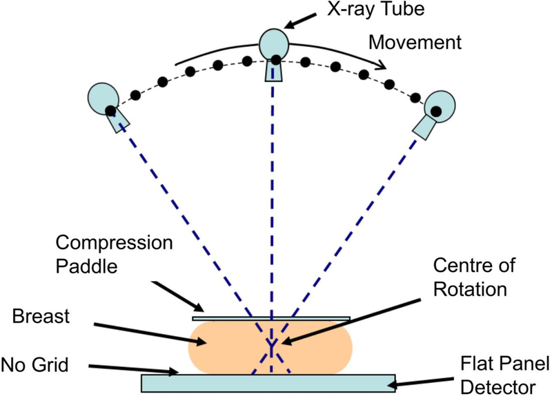
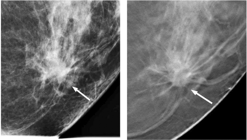
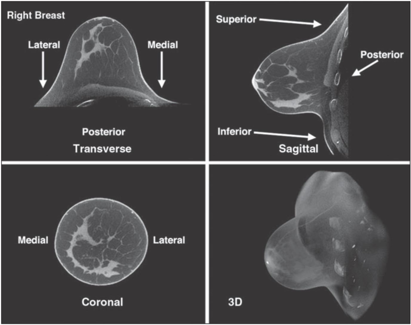
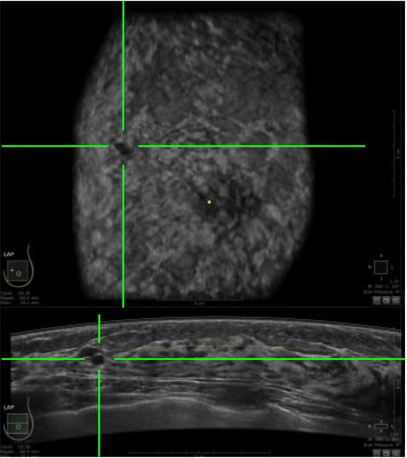
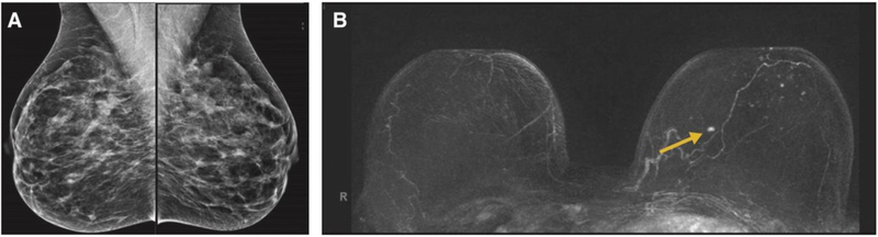
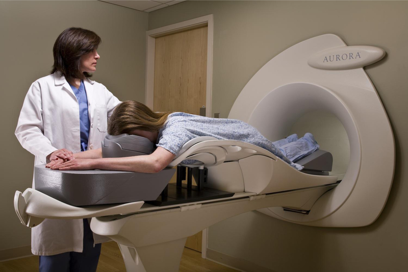
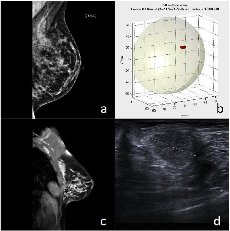
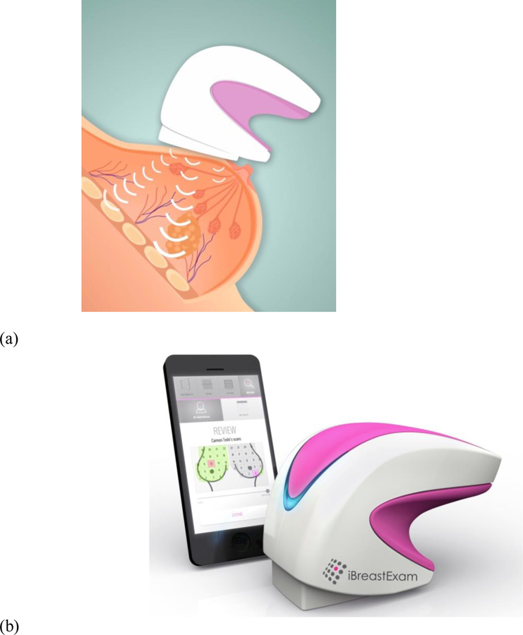
Results: In addition to full-field digital mammography (FFDM), a further nine imaging modalities were identified for review. These included: digital breast tomosynthesis (DBT); breast computed tomography (BCT); automated breast ultrasound (ABUS); fusion of FFDM and ABUS; fusion of DBT and ABUS; magnetic resonance imaging (MRI); optical imaging; radio-wave imaging; and tactile sensor imaging. Important parameters were considered: diagnostic success (sensitivity and specificity), especially in dense breasts; time to acquire the images; and capital cost of the equipment.

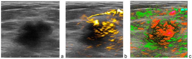
Conclusions: DBT is rapidly replacing FFDM although it still misses invasive cancers in dense tissue. The fusion of ABUS, either with FFDM or DBT, will lead to sensitivity and specificity approaching 100%. The fusion of opto-acoustic imaging with ultrasound holds considerable promise for the future.

Keywords: Breast cancer; Dense breast tissue; Imaging; Screening.


Conflict of interest statement

Competing interests

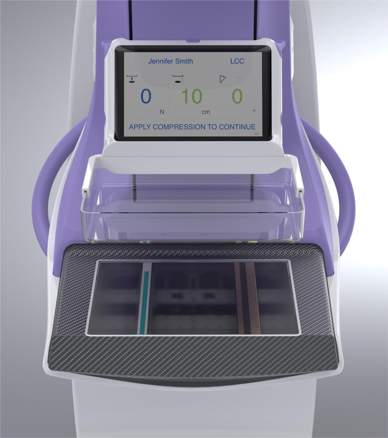
I am an employee of, and a board member and shareholder in CapeRay Medical (Pty) Ltd, a company whose product called Aceso is described in this review paper.

CLV is a board member and shareholder in CapeRay.

Figures

Figure 1
Figure 1
Standard geometry for a digital breast tomosynthesis (DBT) system, illustrating how the X-ray tube moves through an arc while the flat panel detector acquires a series of low dose images of the breast.
Figure 2
Figure 2
A low-grade invasive ductal carcinoma, comparing full-field digital mammography (left) and digital breast tomosynthesis (right). Images courtesy of Dr Steven Poplack.
Figure 3
Figure 3
Breast computer tomography (BCT) images of a 44-year-old woman with heterogeneously dense breasts and normal findings [23]. © American Roentgen Ray Society.
Figure 4
Figure 4
An automated breast ultrasound (ABUS) image in the horizontal plane (bottom) and coronal plane (top), in which the green crosshairs identify the location of a cancerous lesion.
Figure 5
Figure 5
A frontal view of the dual-modality Aceso system that combines full-field digital mammography (FFDM) and automated breast ultrasound (ABUS) in a hermetically-sealed breast platform filled with mineral oil [49]. The ultrasound probe is seen on the left and the digital X-ray camera on the right. Prior to image acquisition, both transducers are located out of the field of view on the right-hand side, and the ABUS and FFDM images are acquired simultaneously as the transducers move from right to left.
Figure 6
Figure 6
Co-registration of the FFDM images in the horizontal plane and the ABUS images in the horizontal, coronal and sagittal planes, as acquired by the Aceso system. (a) A 42-year-old healthy volunteer with extremely dense breast tissue where a benign cyst is highlighted by green crosshairs in the ABUS images, but the lesion is occult in the FFDM image [48]. A 61-year-old patient in which a malignant tumour is clearly co-registered in the ABUS and FFDM images [49]. Note: for the ABUS images, the sagittal plane view is the acquired image, whereas the coronal and horizontal plane views have been reconstructed from the sagittal plane slices.
Figure 7
Figure 7
Fusion of DBT and ABUS with the Siemens Mammomat Inspiration and Acuson S2000 systems [54]. Note: the ultrasound transducer is built into the compression paddle and is located above the breast. © European Society of Radiology.
Figure 8
Figure 8
Fusion of DBT and ABUS images in the sagittal plane, showing co-registration of a cancerous lesion [54]. © European Society of Radiology.
Figure 9
Figure 9
Comparison of FFDM images (A) and MRI images (B) of both breasts of a 45-year-old woman with a moderate history of breast cancer [57]. An invasive ductal carcinoma in the left breast was clearly evident in the MRI image but was occult in the FFDM image. © American Society of Clinical Oncology.
Figure 10
Figure 10
A dedicated 1.5 Tesla breast MRI system manufactured by Aurora Healthcare Corporation [61].
Figure 11
Figure 11
Dual-modality opto-acoustic and ultrasound images of a patient acquired with the Imagio system manufactured by Seno Medical Instruments [66]. (a) A 2.6 cm malignant mass seen on gray-scale ultrasound. (b) Increased haemoglobin due to high density of angiogenesis. (c) Diffuse internal blood deoxygenation [65]. © Alexander Oraevsky.
Figure 12
Figure 12
Patient with a Grade 2 carcinoma [68]. (a) FFDM image which was reported as normal. (b) MARIA image, showing a lesion in the upper outer quadrant (UOQ). (c) Contrast-enhanced MRI, showing a 16 mm irregular mass in the UOQ. (d) Ultrasound image, confirming the lesion. © European Journal of Radiology.
Figure 13
Figure 13
The hand-held iBreastExam [70]. (a) Based on the piezo-electric principle, the 4×4 array of tactile pressure sensors measure the stiffness of the underlying tissues. (b) The device communicates wirelessly with a mobile phone that displays and records the data in real time.

References

    1. Egan RL, “Experience with mammography in a tumor institution. Evaluation of 1,000 studies”, Radiology, 75: 894–900, 1960. - PubMed
    1. Larsson LG, Nyström L, Wall S, Rutqvist L, Andersson I, Bjurstam N, Fagerberg G, Frisell J, Tabár L, “The Swedish randomised mammography screening trials: analysis of their effect on the breast cancer related excess mortality”, Journal of Medical Screening, 3(3) :129–132, 1996. - PubMed
    1. Hendrick RE, Smith RA, Rutledge JH, Smart CR, “Benefit of screening mammography in women aged 40–49: a new meta-analysis of randomized controlled trials”, Journal of the National Cancer Institute Monographs, 22: 87–92, 1997. - PubMed
    1. Pisano ED, “Current status of full-field digital mammography”, Radiology, 214: 26–28, 2000. - PubMed
    1. Kopans DB, “Breast cancer screening: Where have we been and where are we going? A personal perspective based on history, data and experience”, Clinical Imaging, 50: 91–95, 2018. - PubMed

Publication types

MeSH terms