

MHC matching fails to prevent long-term rejection of iPSC-derived neurons in non-human primates
- PMID: 31554807
- PMCID: PMC6761126
- DOI: 10.1038/s41467-019-12324-0
MHC matching fails to prevent long-term rejection of iPSC-derived neurons in non-human primates
Abstract
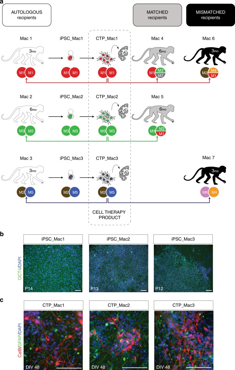
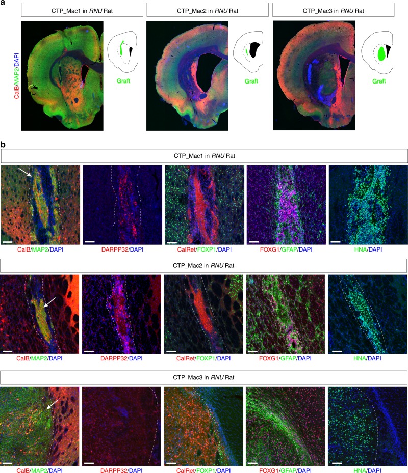
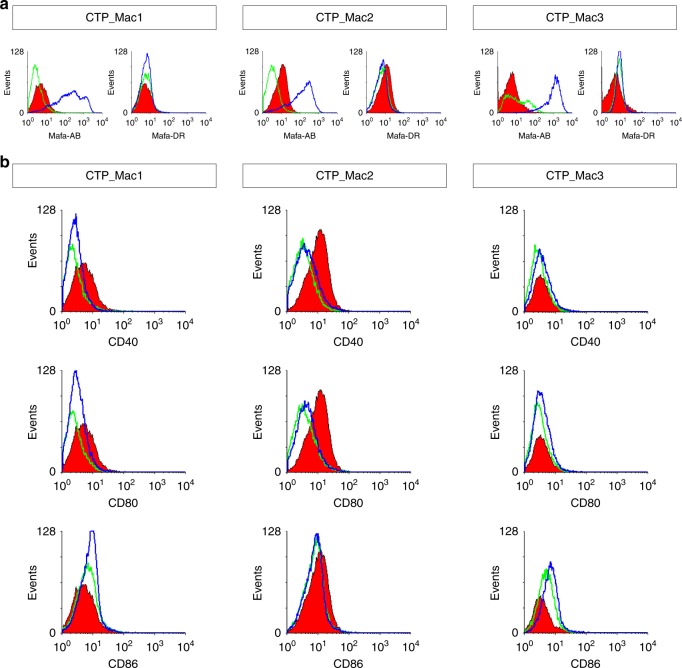
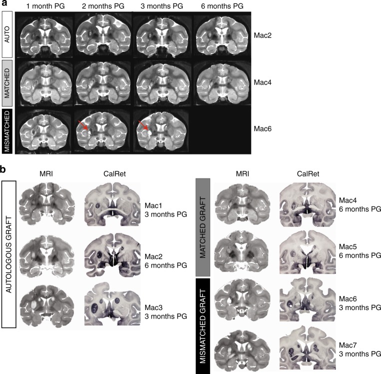
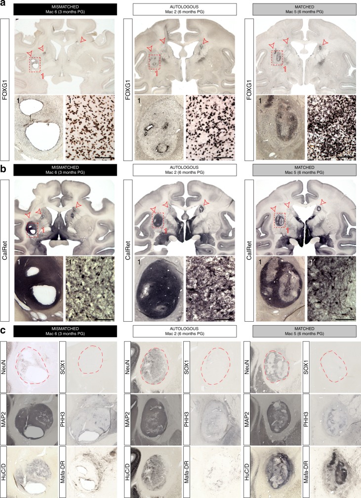
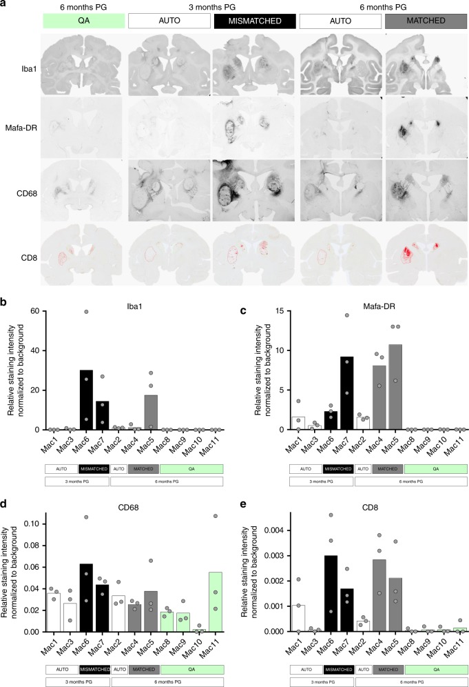
Cell therapy products (CTP) derived from pluripotent stem cells (iPSCs) may constitute a renewable, specifically differentiated source of cells to potentially cure patients with neurodegenerative disorders. However, the immunogenicity of CTP remains a major issue for therapeutic approaches based on transplantation of non-autologous stem cell-derived neural grafts. Despite its considerable side-effects, long-term immunosuppression, appears indispensable to mitigate neuro-inflammation and prevent rejection of allogeneic CTP. Matching iPSC donors' and patients' HLA haplotypes has been proposed as a way to access CTP with enhanced immunological compatibility, ultimately reducing the need for immunosuppression. In the present work, we challenge this paradigm by grafting autologous, MHC-matched and mis-matched neuronal grafts in a primate model of Huntington's disease. Unlike previous reports in unlesioned hosts, we show that in the absence of immunosuppression MHC matching alone is insufficient to grant long-term survival of neuronal grafts in the lesioned brain.
Conflict of interest statement
The authors declare no competing interests.
Figures

Similar articles
-
MHC matching improves engraftment of iPSC-derived neurons in non-human primates.Nat Commun. 2017 Aug 30;8(1):385. doi: 10.1038/s41467-017-00926-5. Nat Commun. 2017. PMID: 28855509 Free PMC article.
-
Direct comparison of autologous and allogeneic transplantation of iPSC-derived neural cells in the brain of a non-human primate.Stem Cell Reports. 2013 Sep 26;1(4):283-92. doi: 10.1016/j.stemcr.2013.08.007. eCollection 2013. Stem Cell Reports. 2013. PMID: 24319664 Free PMC article.
-
Cardiomyocytes Derived from MHC-Homozygous Induced Pluripotent Stem Cells Exhibit Reduced Allogeneic Immunogenicity in MHC-Matched Non-human Primates.Stem Cell Reports. 2016 Mar 8;6(3):312-20. doi: 10.1016/j.stemcr.2016.01.012. Epub 2016 Feb 18. Stem Cell Reports. 2016. PMID: 26905198 Free PMC article.
-
Stem cell transplantation for Huntington's diseases.Methods. 2018 Jan 15;133:104-112. doi: 10.1016/j.ymeth.2017.08.017. Epub 2017 Sep 1. Methods. 2018. PMID: 28867501 Review.
-
[At Home among Strangers: Is It Possible to Create Hypoimmunogenic Pluripotent Stem Cell Lines?].Mol Biol (Mosk). 2019 Sep-Oct;53(5):725-740. doi: 10.1134/S0026898419050045. Mol Biol (Mosk). 2019. PMID: 31661474 Review. Russian.
Cited by
-
Cell therapy for neurological disorders.Nat Med. 2024 Oct;30(10):2756-2770. doi: 10.1038/s41591-024-03281-3. Epub 2024 Oct 15. Nat Med. 2024. PMID: 39407034 Review.
-
Cardiac Cell Therapy with Pluripotent Stem Cell-Derived Cardiomyocytes: What Has Been Done and What Remains to Do?Curr Cardiol Rep. 2022 May;24(5):445-461. doi: 10.1007/s11886-022-01666-9. Epub 2022 Mar 11. Curr Cardiol Rep. 2022. PMID: 35275365 Free PMC article. Review.
-
BioSimia, France CNRS network for nonhuman primate biomedical research in infectiology, immunology, and neuroscience.Curr Res Neurobiol. 2022 Sep 2;3:100051. doi: 10.1016/j.crneur.2022.100051. eCollection 2022. Curr Res Neurobiol. 2022. PMID: 36685763 Free PMC article. Review.
-
Advantages and challenges of using allogeneic vs. autologous sources for neuronal cell replacement in Parkinson's disease: Insights from non-human primate studies.Brain Res Bull. 2025 May;224:111297. doi: 10.1016/j.brainresbull.2025.111297. Epub 2025 Mar 12. Brain Res Bull. 2025. PMID: 40086764 Free PMC article. Review.
-
The Insulin-Producing Cells Generated from Rat Adipose Tissue Mesenchymal Stem Cells via Pdx1 Overexpression Activate an Immune Response both in Vitro and in Vivo.Iran J Med Sci. 2025 Feb 1;50(2):112-123. doi: 10.30476/ijms.2024.101162.3378. eCollection 2025 Feb. Iran J Med Sci. 2025. PMID: 40026296 Free PMC article.
References
Publication types
MeSH terms
LinkOut - more resources
Full Text Sources
Medical
Research Materials
