

A rabbit model for assessing symblepharon after alkali burn of the superior conjunctival sac
- PMID: 31554890
- PMCID: PMC6761117
- DOI: 10.1038/s41598-019-50286-x
A rabbit model for assessing symblepharon after alkali burn of the superior conjunctival sac
Abstract
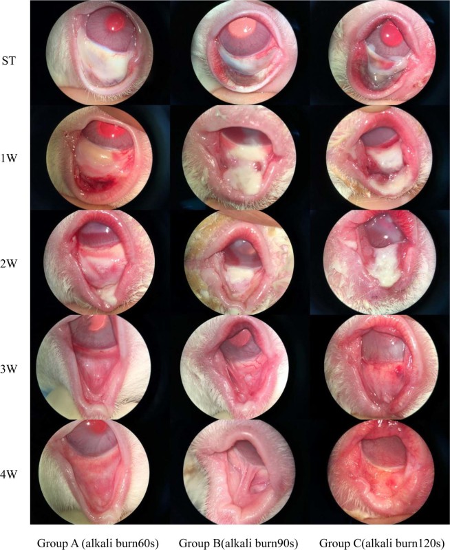
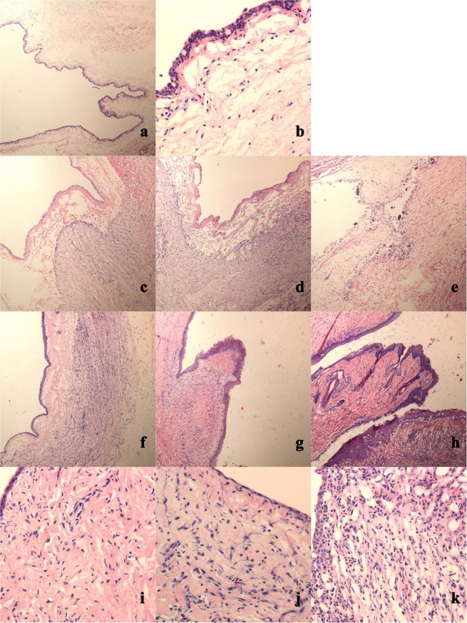
Symblepharon due to chemical burns affects ocular surface health, and there are currently no satisfactory treatments. To improve our understanding of symblepharon, an appropriate animal model is urgently needed. We established a rabbit model of superior conjunctival sac alkaline burn to evaluate symblepharon severity. Alkali burns were induced in rabbits by contacting the superior conjunctival sac with 2 N NaOH-soaked semicircle filter paper (10 mm diameter) for 60 s, 90 s or 120 s. Clinical and histological features were examined, symblepharon severity was evaluated via conjunctival sac depth (grade I - IV) and volume measurements (grade a-d) post-injury at 4 weeks. With increasing alkali burn duration, corneal perforation and symblepharon severity increased. The 60 s group manifested a sub-conjunctiva scar. The 90 s group featured localized adhesion. The 120 s group was characterized by extensive scar hyperplasia and adhesion. The rabbit model exhibited stable and reliable symblepharon following an alkali burn of the superior conjunctival sac. For further research, 90 s is a suitable duration for conjunctival sac burn. The volume measured using conjunctival sac casting was considered when developing a successful evaluation system for symblepharon severity.
Conflict of interest statement
The authors declare no competing interests.
Figures


Similar articles
-
[Experimental study on the effects of chitosan on the conjunctiva scar formation and symblepharon].Zhonghua Yan Ke Za Zhi. 2006 Apr;42(4):313-7. Zhonghua Yan Ke Za Zhi. 2006. PMID: 16762207 Chinese.
-
[Spherical covering foil--silicone implant forming conjunctival fornices].Cesk Slov Oftalmol. 2007 Nov;63(6):396-402. Cesk Slov Oftalmol. 2007. PMID: 18062163 Czech.
-
Gelatin methacryloyl hydrogel eye pad loaded with amniotic extract prevents symblepharon in rabbit eyes.Eur Rev Med Pharmacol Sci. 2020 Oct;24(19):10134-10142. doi: 10.26355/eurrev_202010_23233. Eur Rev Med Pharmacol Sci. 2020. PMID: 33090421
-
Preventing symblepharon formation with a gelatin sponge in the eye of a patient with an alkali burn.Am J Ophthalmol. 1997 Apr;123(4):552-4. doi: 10.1016/s0002-9394(14)70185-0. Am J Ophthalmol. 1997. PMID: 9124256
-
[The ocular surface of severe alkali burns patients on confocal microscopy].Zhonghua Yan Ke Za Zhi. 2010 Jan;46(1):18-24. Zhonghua Yan Ke Za Zhi. 2010. PMID: 20388317 Chinese.
Cited by
-
Sustained release of brimonidine from polydimethylsiloxane-coating silicone rubber implant to reduce intraocular pressure in glaucoma.Regen Biomater. 2023 Apr 24;10:rbad041. doi: 10.1093/rb/rbad041. eCollection 2023. Regen Biomater. 2023. PMID: 37303848 Free PMC article.
-
Molecular mechanisms and treatments for ocular symblephara.Surv Ophthalmol. 2022 Jan-Feb;67(1):19-30. doi: 10.1016/j.survophthal.2021.04.008. Epub 2021 Apr 29. Surv Ophthalmol. 2022. PMID: 33932469 Free PMC article. Review.
-
Risk of Induction of Corneal Neovascularization with Topical Erythropoietin: An Animal Safety Study.J Ophthalmic Vis Res. 2023 Jul 28;18(3):252-259. doi: 10.18502/jovr.v18i3.13772. eCollection 2023 Jul-Sep. J Ophthalmic Vis Res. 2023. PMID: 38737499 Free PMC article.
-
PNP Hydrogel Prevents Formation of Symblephara in Mice After Ocular Alkali Injury.Transl Vis Sci Technol. 2022 Feb 1;11(2):31. doi: 10.1167/tvst.11.2.31. Transl Vis Sci Technol. 2022. PMID: 35191963 Free PMC article.
References
Publication types
MeSH terms
LinkOut - more resources
Full Text Sources
