. 2019 Sep 25;9(1):13903.
doi: 10.1038/s41598-019-50444-1.

Age- and sex-dependent role of osteocytic pannexin1 on bone and muscle mass and strength

Affiliations

Age- and sex-dependent role of osteocytic pannexin1 on bone and muscle mass and strength

Alexandra Aguilar-Perez et al. Sci Rep. .

Abstract

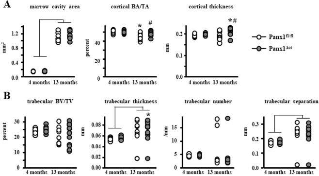
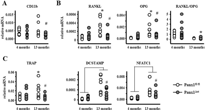
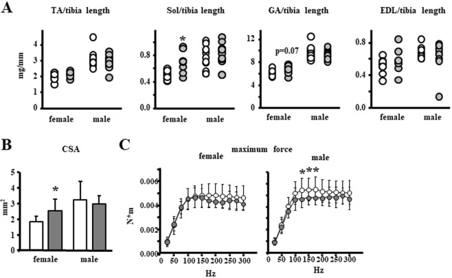
Pannexins (Panxs), glycoproteins that oligomerize to form hemichannels on the cell membrane, are topologically similar to connexins, but do not form cell-to-cell gap junction channels. There are 3 members of the family, 1-3, with Panx1 being the most abundant. All Panxs are expressed in bone, but their role in bone cell biology is not completely understood. We now report that osteocytic Panx1 deletion (Panx1Δot) alters bone mass and strength in female mice. Bone mineral density after reaching skeletal maturity is higher in female Panx1Δot mice than in control Panx1fl/fl mice. Further, osteocytic Panx1 deletion partially prevented aging effects on cortical bone structure and mechanical properties. Young 4-month-old female Panx1Δot mice exhibited increased lean body mass, even though pannexin levels in skeletal muscle were not affected; whereas no difference in lean body mass was detected in male mice. Furthermore, female Panx1-deficient mice exhibited increased muscle mass without changes in strength, whereas Panx1Δot males showed unchanged muscle mass and decreased in vivo maximum plantarflexion torque, indicating reduced muscle strength. Our results suggest that osteocytic Panx1 deletion increases bone mass in young and old female mice and muscle mass in young female mice, but has deleterious effects on muscle strength only in males.


Conflict of interest statement

The authors declare no competing interests.

Figures

Figure 1
Figure 1
Deletion of Panx1 in osteocytes increases bone mass. (A) Panx1 protein levels in calvaria bones from Panx1fl/fl and Panx1Δot mice at 4 months of age were assessed by Western blotting and β-actin was used as a loading control. (B) Representative images of whole body histological preparations (n = 3/genotype) stained by Alcian blue/alizarin red in 6-day-old mice for visual inspection of cartilage (blue) and calcified tissue (magenta). (C) The levels of the apoptosis-related genes were measured by qPCR bone marrow-flushed long bones obtained from female mice. Bars represent mean ± s.d., #p < 0.05 versus Panx1fl/fl mice at the same age, *p < 0.05 versus 4-month-old mice of the same genotype, and lines indicate age effect, by 2-way ANOVA. (D) Body weight and total body, spinal and femoral BMD were assessed monthly in female mice from 1 to 13 months of age by DXA (n = 9–12 per group). ¥p < 0.05 versus Panx1fl/fl mice at the same age by t-test.
Figure 2
Figure 2
Osteocytic Panx1 removal preserves cortical bone volume and increases cortical thickness in aged female mice. (A) Femur cortical bone geometry in the mid-diaphysis was assessed by µCT (n = 9–12 per group). (B) Analysis of cancellous bone microarchitecture in L5 vertebra was assessed by µCT (n = 9–12 per group). Bars represent mean ± s.d., #p < 0.05 versus Panx1fl/fl mice at the same age, *p < 0.05 versus 4-month-old mice of the same genotype, and lines indicate age effect, by 2-way ANOVA.
Figure 3
Figure 3
Aged Panx1Δot female mice have increased mechanical properties in cortical bone. Femur cortical bone structural (A) and material (B) mechanical properties were measured by 3-point bending at 4 and 13 months of age female mice. (n = 9–12 per group). Bars represent mean ± s.d., #p < 0.05 versus Panx1fl/fl mice at the same age, *p < 0.05 versus 4-month-old mice of the same genotype, and lines indicate age effect, by 2-way ANOVA.
Figure 4
Figure 4
Panx1 deletion does not alter bone formation or osteoclast number/surface in cortical bone from female mice. (A) Periosteal (Ps) and endocortical (Ec) MS/BS, MAR, and BFR/BS were measured in unstained femoral cross-sections (n = 9–12 per group). (B) N.Oc/BS, Oc.S/BS, ES/BS were scored on the endocortical surface of the femoral mid-diaphysis after staining for TRAPase/Toluidine blue in Panx1fl/fl and Panx1Δot mice at 4 and 13 months of age (n = 9–12 per group). Bars represent mean ± s.d., Lines indicate age effect, by 2-way ANOVA.
Figure 5
Figure 5
Panx1Δot female mice exhibited minimal changes in vertebral dynamic and static histomorphometric parameters. (A) MS/BS, MAR, and BFR/BS were measure in unstained sections of lumbar vertebra (n = 9–12 per group). (B) N.Oc/BS, Oc.S/BS, ES/BS were scored in vertebral cancellous bone after staining for TRAPase/Toluidine blue in Panx1fl/fl and Panx1Δot mice at 4 and 13 months of age (n = 9–12 per group). (C) N.Ob/BS and Ob.S/BS were scored in lumbar vertebra stained with von Kossa/McNeal (n = 9–12 per group). (D) Circulating markers of bone formation (P1NP, 9–12 per group and ALP, n = 9–12 per group) were measured in serum by ELISA. Bars represent mean ± s.d., #p < 0.05 versus Panx1fl/fl mice at the same age, and lines indicate age effect, by 2-way ANOVA.
Figure 6
Figure 6
Deletion of Panx1 in osteocytes decreases osteoclastic gene expression in aged 13-month-old female mice. (A) CD11b mRNA levels were measured in non-adherent bone marrow cells by qPCR and corrected by GAPDH (n = 9–12 per group). (BD) mRNA levels of RANKL and OPG as well as the indicated osteoclast-related genes were measured in bone marrow-flushed long bones by qPCR and corrected by GAPDH (n = 9–12 per group). Bars represent mean ± s.d., #p < 0.05 versus Panx1fl/fl mice at the same age, *p < 0.05 versus 4-month-old mice of the same genotype, and lines indicate age effect, by 2-way ANOVA.
Figure 7
Figure 7
Panx1Δot female mice exhibit increased muscle mass. (AD) Body weight (A), total lean body mass (B), percent of lean and fat mass (C), and BMD (D) were measured in 10-week-old female and male mice by DXA (Piximus) (n = 9–13 per group). (E) mRNA and protein Panx1 levels were measured in tibialis anterior (TA) muscles by qPCR (corrected by GAPDH, n = 9 and 12 per group), and Western blotting (corrected by β-actin, n = 4 and 3), respectively. Bars represent mean ± s.d., *p < 0.05 versus Panx1fl/fl mice of the same sex, by 2-way ANOVA.
Figure 8
Figure 8
Increased muscle mass in female and decreased muscle strength in male mice lacking Panx1 in osteocytes. (A) Muscle weights normalized by tibia length were measured in 10-week old mice (n = 9–12 per group). TA: tibialis anterior, Sol: soleus, GA: gastrocnemius, EDL: extensor digitorum longus muscles. (B) Soleus physiological cross-sectional area was calculated [CSA = muscle mass (mg)/[(optimal length) × (fiber to muscle length ratio = 0.72) × (density of muscle = 1.06 mg/mm3)]. (C) In vivo assessment of maximum force in males and females (n = 9–13 per group). *p < 0.05 versus Panx1fl/fl mice of the same sex, by t-test.

References

    1. Barbe MT, Monyer H, Bruzzone R. Cell-cell communication beyond connexins: the pannexin channels. Physiology. (Bethesda.) 2006;21:103–114. - PubMed
    1. Penuela S, et al. Pannexin 1 and pannexin 3 are glycoproteins that exhibit many distinct characteristics from the connexin family of gap junction proteins. J. Cell Sci. 2007;120:3772–3783. doi: 10.1242/jcs.009514. - DOI - PubMed
    1. Penuela S, Gehi R, Laird DW. The biochemistry and function of pannexin channels. Biochim. Biophys. Acta. 2013;1828:15–22. doi: 10.1016/j.bbamem.2012.01.017. - DOI - PubMed
    1. Penuela S, Harland L, Simek J, Laird DW. Pannexin channels and their links to human disease. Biochem. J. 2014;461:371–381. doi: 10.1042/BJ20140447. - DOI - PubMed
    1. Sosinsky GE, et al. Pannexin channels are not gap junction hemichannels. Channels (Austin.) 2011;5:193–197. doi: 10.4161/chan.5.3.15765. - DOI - PMC - PubMed

Publication types

LinkOut - more resources