. 2019 Sep 4:10:959.
doi: 10.3389/fphar.2019.00959. eCollection 2019.

Antiviral Properties of R. tanguticum Nanoparticles on Herpes Simplex Virus Type I In Vitro and In Vivo

Affiliations

Antiviral Properties of R. tanguticum Nanoparticles on Herpes Simplex Virus Type I In Vitro and In Vivo

Meng-Xin Shen et al. Front Pharmacol. .

Abstract

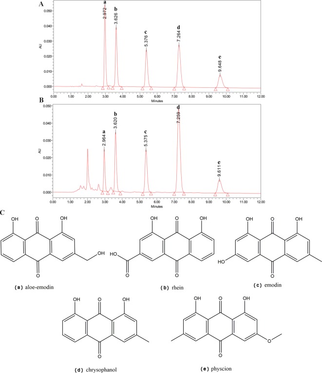
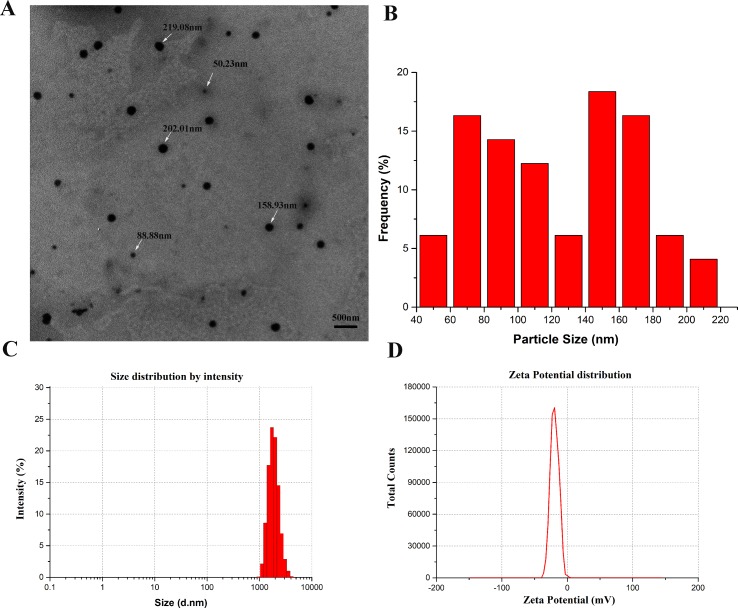
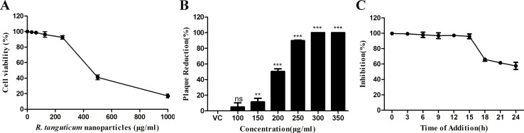
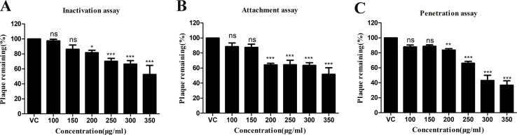
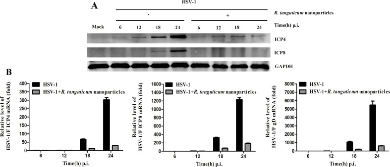
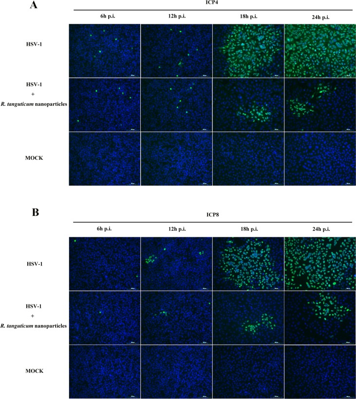
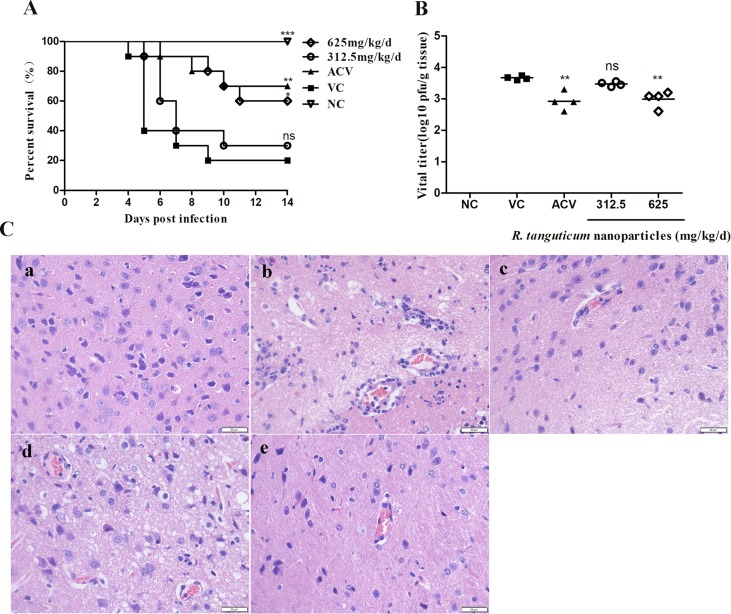
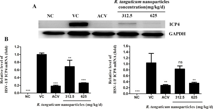
Herpes simplex virus type 1 (HSV-1), an enveloped DNA virus, plays a key role in varieties of diseases including recurrent cold sores, keratoconjunctivitis, genital herpes and encephalitis in humans. Great efforts have been made in developing more effective and less side-effects anti-herpes simplex virus agents, including traditional Chinese herbal medicines. In the present study, we evaluated the antiviral efficacy of Rheum tanguticum nanoparticles against HSV-1 in vitro and in vivo. R. tanguticum nanoparticles could inactivate the HSV-1 virions and block the viral attachment and entry into cells. Time-of-addition assay indicated that R. tanguticum nanoparticles could interfere with the entire phase of viral replication. Besides, R. tanguticum nanoparticles showed the ability to inhibit the mRNA expression of HSV-1 immediate early gene ICP4 and early gene ICP8 as well as the expression of viral protein ICP4 and ICP8. Moreover, R. tanguticum nanoparticles have been proved to protect mice against HSV-1 induced lethality by decreasing the viral load and alleviated pathological changes in brain tissues. In conclusion, we demonstrated that R. tanguticum nanoparticles could inhibit HSV-1 infection through multiple mechanisms. These results suggest that R. tanguticum nanoparticles may have novel roles in the treatment of HSV-1 infection.

Keywords: HSV-1; R. tanguticum nanoparticles; antiviral; mouse model; viral protein.


Figures

Figure 1
Figure 1
HPLC analysis of R. tanguticum nanoparticles. (A) HPLC of the standard reference compounds. (B) The chemical contents of R. tanguticum nanoparticles, peak a for aloe–emodin, peak b for rhein, peak c for emodin, peak d for chrysophanol, and peak e for physcion, respectively. (C) Structure of identified components of R. tanguticum nanoparticles.
Figure 2
Figure 2
Characterization of R. tanguticum nanoparticles (A) Transmission electron microscopy (TEM) images of R. tanguticum nanoparticles. (B) TEM size distribution histogram. (C) Dynamic light scattering (DLS) size distribution histogram. (D) Zeta potential distribution of R. tanguticum nanoparticles.
Figure 3
Figure 3
R. tanguticum nanoparticles inhibition of HSV-1 replication. (A) HEp-2 cells in 96-well plates were treated with serial dilution of R. tanguticum nanoparticles for 72 h and the viability were measured by the MTT assay. (B) Inhibitory effects of R. tanguticum nanoparticles on HSV-1 infection. HEp-2 cells infected with HSV-1 were treated with different concentrations of R. tanguticum nanoparticles and were subjected to the plaque reduction assay. (C) Time-of-addition assay. HEp-2 cells were infected with HSV-1 and then treated with R. tanguticum nanoparticles (350 µg/ml) at indicated intervals. The progeny virus yields were determined by plaque assay. Values are represented as the mean ± standard deviation of three individual experiments. “VC” is the abbreviation of the “virus control group”.*p < 0.05; **p < 0.01; ***p < 0.001; n.s., not significant.
Figure 4
Figure 4
The anti-inactivation (A) anti-attachment (B) and anti-penetration (C) activities of R. tanguticum nanoparticles. Values are represented as the mean ± standard deviation of three individual experiments. “VC” is the abbreviation of the “virus control group”.*p < 0.05; **p < 0.01; ***p < 0.001; n.s., not significant.
Figure 5
Figure 5
The effect of R. tanguticum nanoparticles on HSV-1 immediate-early and early genes expression. (A) Western blot analysis: HEp-2 cells were infected with HSV-1 at an MOI of 0.01 and then treated with or without 350 µg/ml R. tanguticum nanoparticles. The cells were harvested at each time point (6, 12, 18, and 24 h) for western blot analysis of ICP4 and ICP8. (B) Real-time quantitative PCR analysis: HSV-1 (MOI = 0.01) infected cells were treated with 350 µg/ml R. tanguticum nanoparticles for 6, 12, 18, and 24 h.p.i. post-infection. Total RNA was extracted and subjected to cDNA synthesis. The real-time quantitative PCR was performed ICP4-, ICP8-, and gD-specific primers. Values are represented as the mean ± standard deviation of three individual experiments.
Figure 6
Figure 6
Immunofluorescence assay of HSV-1 infected cells treated with R. tanguticum nanoparticles. (A–B) HEp-2 cells were infected with HSV-1 at an MOI of 0.01 and incubated with 350 µg/ml R. tanguticum nanoparticles. At 6, 12, 18, and 24 h.p.i., cells were fixed with paraformaldehyde and blocked in 1% BSA. After blocking, cells were stained of ICP4 and ICP8. The nucleus was stained with DAPI and the green foci to indicate the presence of HSV-1protein.
Figure 7
Figure 7
R. tanguticum nanoparticles alleviated HSV-1 infection in mice. Kunming mice (n = 10 mice/group) infected with 5 LD50 of HSV-1 were orally administered with 625 mg/kg/day and 312.5 mg/kg/day R. tanguticum nanoparticles, respectively. A 0.9% saline was used in viral control and normal control group. (A) The survival rates were collected daily for 14 day. (B) Brain Virus titers were determined by plaque assay at the 5th d.p.i. (C) Histological observations of cerebral tissues for mice sacrificed at the 5th d.p.i. Scale bar = 20 µm. (a) Mock infected mice treated with 0.9% saline (normal control, NC); (b) HSV-1-infected mice treated with 0.9% saline (viral control); (c) HSV-1-infected mice treated with acyclovir; (d–e) HSV-1-infected mice treated with 312.5 or 625 mg/kg/day of R. tanguticum nanoparticles.*p < 0.05; **p < 0.01; ***p < 0.001; n.s., not significant.
Figure 8
Figure 8
R. tanguticum nanoparticles treatment reduced the genes and protein expression in HSV-1 infected mice. Kunming mice (n = 4 mice/group) infected with 5 LD50 of HSV-1 were orally administered with 625 mg/kg/day, 312.5 mg/kg/day R. tanguticum nanoparticles, respectively. A 0.9% saline was used in viral control and normal control group. The mice were scheduled for sacrifice at 5 d.p.i. (A) The expression of the ICP4 protein of the brain tissue was detected by Western blot analysis. (B) The mRNA levels of the ICP4 and ICP8 in treated and control groups were determined by real-time quantitative PCR analysis.*p < 0.05; **p < 0.01; ***p < 0.001; n.s., not significant.

References

    1. Arokiyaraj S., Vincent S., Saravanan M., Lee Y., Oh Y. K., Kim K. H. (2017). Green synthesis of silver nanoparticles using Rheum palmatum root extract and their antibacterial activity against Staphylococcus aureus and Pseudomonas aeruginosa. Artif. Cells Nanomed. Biotechnol. 45 (2), 372–379. 10.3109/21691401.2016.1160403 - DOI - PubMed
    1. Ben Salem A. N., Zyed R., Lassoued M. A., Nidhal S., Sfar S., Mahjoub A. (2012). Plant-Derived Nanoparticles Enhance Antiviral Activity against Coxsakievirus B3 by Acting on Virus Particles and Vero Cells. Dig. J. Nanomater. Bios. 7 (2), 737–744.
    1. Bertol J. W., Rigotto C., de Padua R. M., Kreis W., Barardi C. R. M., Braga F. C., et al. (2011). Antiherpes activity of glucoevatromonoside, a cardenolide isolated from a brazilian cultivar of digitalis lanata. Antivir. Res. 92 (1), 73–80. 10.1016/j.antiviral.2011.06.015 - DOI - PubMed
    1. Burcea M., Gheorghe A., Pop M. (2015). Incidence of herpes simplex virus keratitis in HIV/AIDS patients compared with the general population. J. Med. Life 8 (1), 62–63. - PMC - PubMed
    1. Cagno V., Andreozzi P., D’Alicarnasso M., Silva P. J., Mueller M., Galloux M., et al. (2018). Broad-spectrum non-toxic antiviral nanoparticles with a virucidal inhibition mechanism. Nature Materials 17 (2), 195–19+. 10.1038/nmat5053 - DOI - PubMed

LinkOut - more resources