. 2019 Aug 21:10:1962.
doi: 10.3389/fimmu.2019.01962. eCollection 2019.

Sensitization of Tumors for Attack by Virus-Specific CD8+ T-Cells Through Antibody-Mediated Delivery of Immunogenic T-Cell Epitopes

Affiliations

Sensitization of Tumors for Attack by Virus-Specific CD8+ T-Cells Through Antibody-Mediated Delivery of Immunogenic T-Cell Epitopes

Julian P Sefrin et al. Front Immunol. .

Abstract

Anti-tumor immunity is limited by a number of factors including the lack of fully activated T-cells, insufficient antigenic stimulation and the immune-suppressive tumor microenvironment. We addressed these hurdles by developing a novel class of immunoconjugates, Antibody-Targeted Pathogen-derived Peptides (ATPPs), which were designed to efficiently deliver viral T-cell epitopes to tumors with the aim of redirecting virus-specific memory T-cells against the tumor. ATPPs were generated through covalent binding of mature MHC class I peptides to antibodies specific for cell surface-expressed tumor antigens that mediate immunoconjugate internalization. By means of a cleavable linker, the peptides are released in the endosomal compartment, from which they are loaded into MHC class I without the need for further processing. Pulsing of tumor cells with ATPPs was found to sensitize these for recognition by virus-specific CD8+ T-cells with much greater efficiency than exogenous loading with free peptides. Systemic injection of ATPPs into tumor-bearing mice enhanced the recruitment of virus-specific T-cells into the tumor and, when combined with immune checkpoint blockade, suppressed tumor growth. Our data thereby demonstrate the potential of ATPPs as a means of kick-starting the immune response against "cold" tumors and increasing the efficacy of checkpoint inhibitors.

Keywords: CD8+ T-cell; antibody-targeted pathogen-derived peptides (ATPP); antigen-armed antibodies; cancer immunotherapy; immune tolerance; immunogenicity; targeted therapy.


Figures

Figure 1
Figure 1
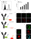
Structure and proposed mode of action of ATPP immunoconjugates. (A) Representative structure of ATPP immunoconjugates. Free lysine residues on the antibody serve to attach an SPDP linker (red) via an amide bond. The immunogenic peptide is connected to the linker via a cysteine using a disulfide bond. The cysteine can reside in the middle, on the C- or N-terminus of the peptide. (B) Proposed model for the mode of action of ATPPs. A: Binding of ATPP to cell surface expressed target antigen. B: Internalization of ATPP into endosomal compartment. C: Release of T-cell response eliciting peptide from the immunoconjugate in the endosomal compartment. D: Loading of MHC-I molecules with released peptide. E: Routing of peptide-loaded MHC-I molecules to target cell surface. F: Recognition of peptide-loaded MHC-I molecules on target cell surface by peptide-specific CD8+ cytotoxic T-cells.
Figure 2
Figure 2
Peptide release from ATPP occurs following internalization in endosomes. (A) Binding and (B) internalization of αCDCP1 Ab and αCDCP1-EBV_1 ATPP on MDA-MB231 cells as determined by flow cytometry. (C) Graphical representation of the αCDCP1 FRET disulfide reporter conjugate. Excitation of the donor chromophore (BODIPY) triggers FRET to the acceptor chromophore (Rhodamine, red signal, upper panel). Release of the acceptor upon disulfide reduction results in green (BODIPY) signals (lower panel). (D) Confocal time-lapse imaging of MDA-MB231 cells using the αCDCP1 FRET disulfide reporter conjugate. (E) Illustration of separate excitation of donor (BODIPY) and acceptor (Rhodamine) in the cleaved αCDCP1 FRET disulfide reporter conjugate. (F) Magnification of an endosome using separate excitation of BODIPY and Rhodamine. (G) Magnification of an endosome upon co-staining with αIgG antibody. Scale bars: 3 μm.
Figure 3
Figure 3
ATPP loaded tumor cells activate peptide-specific CD8+ T-cells. (A) Activation of peptide-specific CD8+ T-cells as measured by IFNγ ELISA after treating indicated CDCP1+, HLA-matched cancer cell lines with αCDCP1-EBV_1, (B) αCDCP1-EBV_2, or (C) αCDCP1-FLU ATPP. Tumor cells exogenously loaded with free peptides (pEBV_1, pEBV_2, pFLU) serve as positive control and unconjugated αCDCP1 Ab as negative control. T-cells were added after 24 h and culture supernatant was harvested after additional 24 h of incubation. (D) In a similar manner, CD138-expresssing U266B1 cells were treated with αCD138-EBV_1 ATPP. For each chart, data represent triplicate values and error bars indicate standard deviation. Experiments have been reproduced with 6 different cancer cell lines using 5 different T-cell donors. For additional data please refer to Supplementary Figure 2.
Figure 4
Figure 4
ATPP-mediated peptide delivery is Ab target-dependent and bypasses the antigen processing machinery. Activation of peptide-specific CD8+ T-cells was measured by IFNγ ELISA after co-incubation with CDCP1+, HLA-matched cancer cells that were pre-treated with αCDCP1-EBV_1 ATPP, other ATPPs or synthetic peptide as indicated. (A) Accessibility of MHC class I was blocked by addition of pan-HLA class I binding antibody W6/32 at indicated concentrations. (B) Sensitization for T-cell recognition by αCDCP1-EBV_1 ATPP was compared with that by an ATPP in which the peptide was bound through a non-cleavable thioether linker, as well as by ATPPs targeting the CD22 or CD79b antigens, which are not expressed on the tumor cells. All compounds were used at 0.132 nM. (C) Comparison of T-cell recognition of target cells that were pre-treated with αCDCP1-EBV_1 ATPP comprising the mature (minimal) T-cell epitope, an ATPP comprising an N-terminally extended version of this epitope (EBV_1_long), or the synthetic equivalents of these peptides at concentrations indicated. For each chart, data represent triplicate values and error bars indicate standard deviation. MHC dependency has been shown in 6 (2 donors, 2 cell lines), target dependency in 7 (3 donors, 2 cell lines) and independency of Ag processing in 2 different experiments (2 donors, 2 cell lines). For additional data please refer to Supplementary Figures 2H, 3.
Figure 5
Figure 5
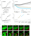
Selective T-cell mediated killing of ATPP-treated tumor cells in vitro. (A) Killing of CDCP1+, HLA-A02:01+ MDA-MB231 cells by pEBV_1 specific CD8+ T-cells at varying effector-to-target ratios after incubation with αCDCP1-EBV_1, non-targeting αCD22-EBV_1 ATPP or free peptide (pEBV_1). Percentage of lysis was determined by LDH quantification in the supernatant after 24 h. (B) Real-time analysis of target cell killing in the xCELLigence system using CDCP1+, HLA-A02:01+ HCT-116 cells, which were pre-incubated for 24 h with indicated ATPPs or synthetic peptides. pEBV_1-specific CD8+ T-cells were added at t = 0 at an effector-to-target ratio of 3:1. (C) Percentage of tumor cell killing was calculated with data received from (B) at indicated time-points. (D) Time-lapse microscopy of co-cultures of CMFDA-labeled HCT-116 cells (green) and PKH-26-labeled pEBV_1-specific CD8+ T-cells (red). HCT-116 were pre-treated with 0.132 nM αCDCP1-EBV_1 ATPP or control αCD22-EBV_1 ATPP. T-cells were added at an effector-to-target ratio of 2:1. For each chart, data represent triplicate values and error bars indicate standard deviation. ATPP mediated killing of target cells vs. non-targeting and non-cleavable constructs has been shown in 4 (2 donors, 2 cell lines) for LDH release and in 13 different experiments (3 donors, 2 cell lines) employing the xCELLigence system.
Figure 6
Figure 6
ATPPs can mediate IFNγ secretion and killing by freshly isolated memory CD8+ T-cells. (A) Flow cytometric analysis of freshly isolated CD8+ T-cells for the frequency of pEBV_1-specific T-cells by means of pentamer staining. (B) Activation of freshly isolated CD8+ T-cells as measured by IFNγ ELISA by αCDCP1-EBV_1 ATPP-loaded MDA-MB231 tumor cells. Free peptide (pEBV_1) serves as reference and non-cleavable αCDCP1- as well as non-targeting αCD22- and αCD79b-ATPP as controls. (C) Killing of MDA-MB231 tumor cells by freshly isolated CD8+ T-cells after pre-treatment with indicated ATPPs or synthetic peptides. Total CD8+ T-cell-to-target ratio was 20:1, meaning that pEBV_1-specific CD8+ T-cell-to-target ratio was ~1:10. For each chart, data represent triplicate values and error bars indicate standard deviation.
Figure 7
Figure 7
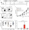
ATPPs efficiently recruit peptide-specific T-cells into the tumor and mediate suppression of tumor growth in vivo. (A) Study outline, using the CDCP1+, HLA-A02:01+ MDA-MB231 s.c. breast cancer xenograft model in NOG mice and adoptive transfer of in vitro-expanded pEBV_1-specific human CD8+ T-cells. (B) Flow cytometric phenotyping of T-cells prior to transfer regarding peptide specificity, CD4, CD8, and PD1 expression. (C) Analysis of target (CDCP1) and PD-L1 expression in s.c. MDA-MB231 tumors by flow cytometry. (D) Kinetics of MDA-MB231 tumor growth as determined by caliper measurement. Mice were either only injected s.c. with tumor cells (Tumor control), additionally received i.v. T-cells (T-cell control) and/or were treated with 20 mg/kg/week αCDCP1-EBV_1 ATPP, αCDCP1 antibody (Ab), and/or 5 mg/kg/week αPD1 Ab. (E) Endpoint analysis of tumor volume on day 39. (F) Analysis of pEBV_1-specific CD8+ tumor infiltrating lymphocytes (TILs) in s.c. MDA-MB231 tumors after study termination. Data was acquired by means of flow cytometry using peptide-MHC pentamers. For each chart, data is shown as mean and error bars indicate standard error of mean (n = 10). The p-values represent comparisons between groups using one-way ANOVA followed by Tukey's multiple comparison test. **p < 0.01, ***p < 0.001, ****p < 0.0001.

References

    1. Ribas A, Wolchok JD. Cancer immunotherapy using checkpoint blockade. Science. (2018) 359:1350–5. 10.1126/science.aar4060 - DOI - PMC - PubMed
    1. Neefjes J, Jongsma MLM, Paul P, Bakke O. Towards a systems understanding of MHC class I and MHC class II antigen presentation. Nat Rev Immunol. (2011) 11:823–36. 10.1038/nri3084 - DOI - PubMed
    1. DiJoseph JF, Dougher MM, Kalyandrug LB, Armellino DC, Boghaert ER, Hamann PR, et al. . Antitumor efficacy of a combination of CMC-544 (inotuzumab ozogamicin), a CD22-targeted cytotoxic immunoconjugate of calicheamicin, and rituximab against non-Hodgkin's B-cell lymphoma. Clin Cancer Res. (2006) 12:242–9. 10.1158/1078-0432.CCR-05-1905 - DOI - PubMed
    1. Dornan D, Bennett F, Chen Y, Dennis M, Eaton D, Elkins K, et al. . Therapeutic potential of an anti-CD79b antibody-drug conjugate, anti-CD79b-vc-MMAE, for the treatment of non-Hodgkin lymphoma. Blood. (2009) 114:2721–9. 10.1182/blood-2009-02-205500 - DOI - PubMed
    1. Ikeda H, Hideshima T, Fulciniti M, Lutz RJ, Yasui H, Okawa Y, et al. . The monoclonal antibody nBT062 conjugated to cytotoxic Maytansinoids has selective cytotoxicity against CD138-positive multiple myeloma cells in vitro and in vivo. Clin Cancer Res. (2009) 15:4028–37. 10.1158/1078-0432.CCR-08-2867 - DOI - PubMed

Publication types