Novel Mutations Associated With Various Types of Corneal Dystrophies in a Han Chinese Population
- PMID: 31555324
- PMCID: PMC6726741
- DOI: 10.3389/fgene.2019.00881
Novel Mutations Associated With Various Types of Corneal Dystrophies in a Han Chinese Population
Abstract
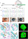
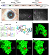
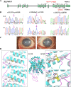
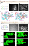
Aims: To study the genetic spectra of corneal dystrophies (CDs) in Han Chinese patients using next-generation sequencing (NGS). Methods: NGS-based targeted region sequencing was performed to evaluate 71 CD patients of Han Chinese ethnicity. A custom-made capture panel was designed to capture all coding exons and untranslated regions plus 25 bp of intronic flanking sequences of 801 candidate genes for eye diseases. The Genome Analysis Tool Kit Best Practices pipeline and an intensive computational prediction pipeline were applied for the analysis of pathogenic variants. Results: We achieved a mutation detection rate of 59.2% by NGS. Eighteen known mutations in CD-related genes were found in 42 out of 71 patients, and these cases showed a genotype-phenotype correlation consistent with previous reports. Nine novel variants that were likely pathogenic were found in various genes, including CHST6, TGFBI, SLC4A11, AGBL1, and COL17A1. These variants were all predicted to be protein-damaging by an intensive computational analysis. Conclusions: This study expands the spectra of genetic mutations associated with various types of CDs in the Chinese population and highlights the clinical utility of targeted NGS for genetically heterogeneous CD.
Keywords: Han Chinese population; corneal dystrophies; mutations; next-generation sequencing; targeted-region sequencing.
Figures


Similar articles
-
Evaluation of the Genetic Variation Spectrum Related to Corneal Dystrophy in a Large Cohort.Front Cell Dev Biol. 2021 Mar 18;9:632946. doi: 10.3389/fcell.2021.632946. eCollection 2021. Front Cell Dev Biol. 2021. PMID: 33816482 Free PMC article.
-
Clinical and genetic features of TGFBI-linked corneal dystrophies in Mexican population: description of novel mutations and novel genotype-phenotype correlations.Exp Eye Res. 2009 Aug;89(2):172-7. doi: 10.1016/j.exer.2009.03.004. Epub 2009 Mar 18. Exp Eye Res. 2009. PMID: 19303004
-
Variant Landscape of 15 Genes Involved in Corneal Dystrophies: Report of 30 Families and Comprehensive Analysis of the Literature.Int J Mol Sci. 2023 Mar 6;24(5):5012. doi: 10.3390/ijms24055012. Int J Mol Sci. 2023. PMID: 36902444 Free PMC article.
-
Genetic analysis of CHST6 and TGFBI in Turkish patients with corneal dystrophies: Five novel variations in CHST6.Mol Vis. 2016 Oct 26;22:1267-1279. eCollection 2016. Mol Vis. 2016. PMID: 27829782 Free PMC article.
-
Clinical and genetic characteristics of Leber congenital amaurosis with novel mutations in known genes based on a Chinese eastern coast Han population.Graefes Arch Clin Exp Ophthalmol. 2016 Nov;254(11):2227-2238. doi: 10.1007/s00417-016-3428-5. Epub 2016 Jul 16. Graefes Arch Clin Exp Ophthalmol. 2016. PMID: 27422788
Cited by
-
Investigation of the functional impact of CHED- and FECD4-associated SLC4A11 mutations in human corneal endothelial cells.PLoS One. 2024 Jan 22;19(1):e0296928. doi: 10.1371/journal.pone.0296928. eCollection 2024. PLoS One. 2024. PMID: 38252645 Free PMC article.
-
Novel Mutations in COL6A3 That Associated With Peters' Anomaly Caused Abnormal Intracellular Protein Retention and Decreased Cellular Resistance to Oxidative Stress.Front Cell Dev Biol. 2020 Nov 10;8:531986. doi: 10.3389/fcell.2020.531986. eCollection 2020. Front Cell Dev Biol. 2020. PMID: 33304895 Free PMC article.
-
From Genes to Disease: Reassessing LOXHD1 and AGBL1's Contribution to Fuchs' Dystrophy.Int J Mol Sci. 2025 Apr 3;26(7):3343. doi: 10.3390/ijms26073343. Int J Mol Sci. 2025. PMID: 40244234 Free PMC article.
-
Genetic mutations and molecular mechanisms of Fuchs endothelial corneal dystrophy.Eye Vis (Lond). 2021 Jun 15;8(1):24. doi: 10.1186/s40662-021-00246-2. Eye Vis (Lond). 2021. PMID: 34130750 Free PMC article. Review.
-
SLC4A11 Revisited: Isoforms, Expression, Functions, and Unresolved Questions.Biomolecules. 2025 Jun 16;15(6):875. doi: 10.3390/biom15060875. Biomolecules. 2025. PMID: 40563515 Free PMC article. Review.
References
LinkOut - more resources
Full Text Sources
Miscellaneous

