. 2019 Aug;8(4):500-512.
doi: 10.21037/tlcr.2019.07.01.

Influence and mechanism of lung cavitation development on antiangiogenic therapy

Affiliations

Influence and mechanism of lung cavitation development on antiangiogenic therapy

Man Jiang et al. Transl Lung Cancer Res. 2019 Aug.

Abstract

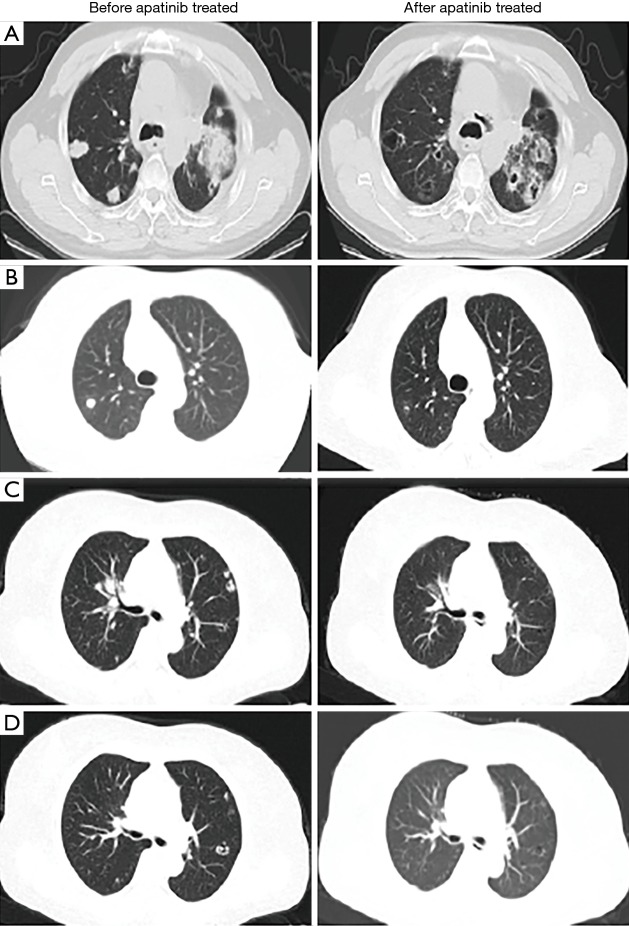
Background: Antiangiogenic agent-treated patients usually develop cavitation in their lung lesions. The clinical significance of lung cavitation development during antiangiogenic therapy has not been determined yet. Herein, we evaluated the clinical outcomes of patients who developed tumor cavitation following apatinib treatment and explored the mechanisms.

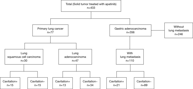
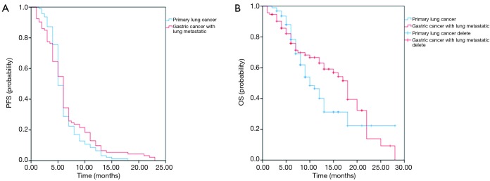
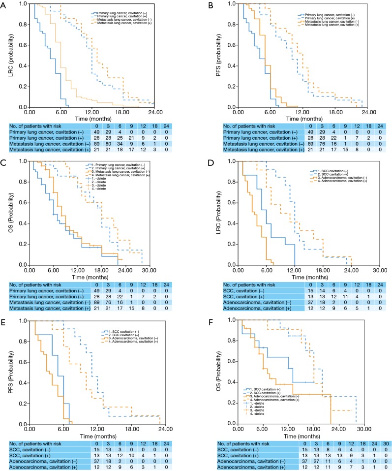
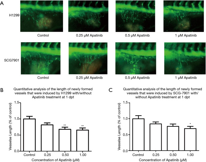
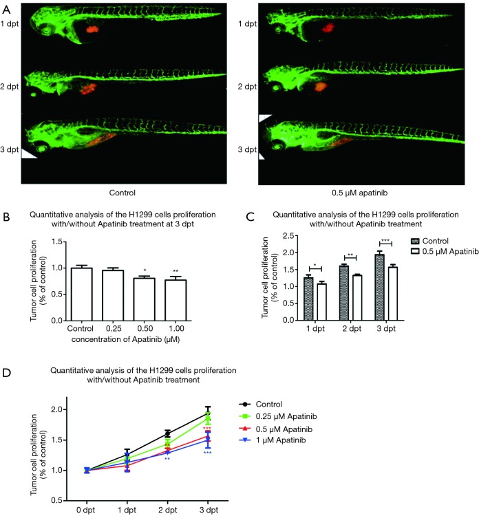
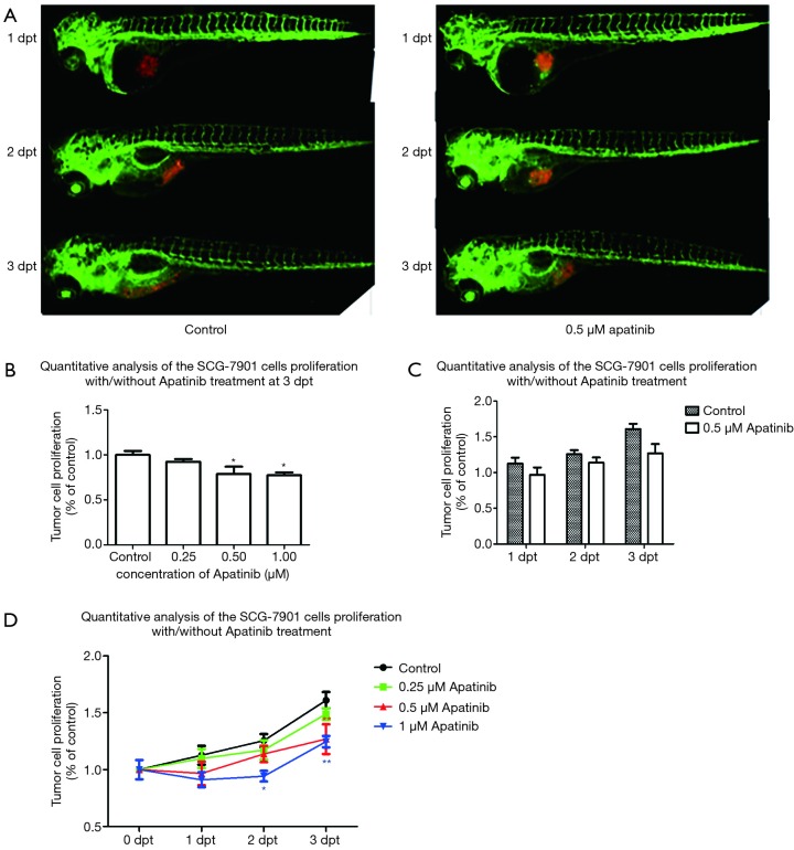
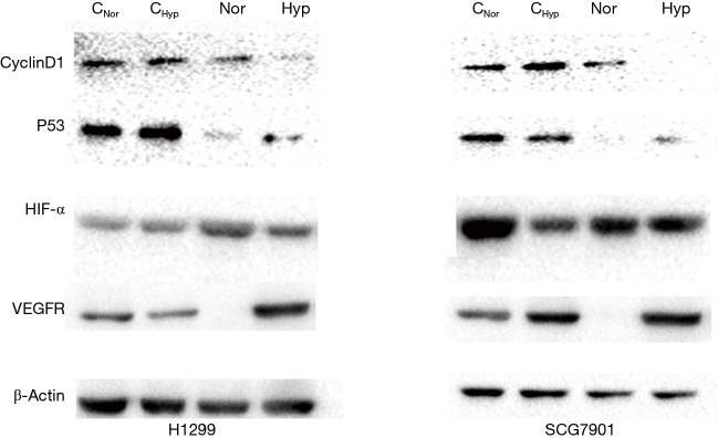
Methods: In this study (Clinical Trial No. NCT03629691), 187 patients (77 lung cancer and 110 gastric adenocarcinoma patients) who had progressed or relapsed after undergoing at least two lines of systemic therapy in accordance with the NCCN guidelines for primary or metastatic lung tumors were treated with apatinib at a dosage of 250 mg per day between February 1, 2015 and May 19, 2017. The effect of lung cavitation development on locoregional control (LRC), progression-free survival (PFS), and overall survival (OS) was analyzed with Kaplan-Meier estimates and compared with the log-rank test. Zebrafish experiments were used to study the anticancer mechanism of apatinib in different tumors. Western-blotting was used to analyze the expression of Cyclin D1, p53, HIF-α, and VEGFR before and after apatinib treatment in both normoxia and hypoxia.

Results: Cavitation development was beneficial in patients receiving apatinib therapy regardless of whether they had primary or metastatic lung cancer. Zebrafish experiments showed that apatinib inhibited tumor growth by both suppressing vascular growth and inhibiting cell proliferation. Vascular proliferation induced by the H1299 cell lines showed higher sensitivity to apatinib than that induced by the SCG-7901 cell line. However, apatinib showed weak tumor type selectivity on cell proliferation inhibition in vivo. Under hypoxic conditions, apatinib could not inhibit the protein expression of VEGFR and HIF-α in both cell lines; however, apatinib decreased the expression of cyclin D1 and P53 significantly.

Conclusions: Lung cavitation development is common with apatinib therapy and is a potential prognostic marker. Apatinib inhibits tumor growth by both vessel growth inhibition and proliferation inhibition.

Keywords: Apatinib; anticancer mechanism; hypoxia; lung cavitation development; progression-free survival (PFS).


Conflict of interest statement

Conflicts of Interest: The authors have no conflicts of interest to declare.

Figures

Figure 1
Figure 1
Trial profile.
Figure 2
Figure 2
Chest CT scans of patients. (A) A 60-year-old man with stage IV lung squamous cell carcinoma treated with apatinib. His baseline chest CT scan prior to therapy demonstrated a solid dominant mass in the left lung, and four nodules were noted in the right lower lobe. A follow-up CT scan at 1.5 months after apatinib therapy demonstrated that a cavity developed in the left lung within the dominant mass, decreasing in the size of the mass. The four nodules in the right lung developed cavitations. (B) A 60-year-old woman with stage IV lung adenocarcinoma who underwent right lower lobectomy 4 years ago, presenting with histologically confirmed recurrent disease in the right lung nodules. Her baseline chest CT scan prior to therapy demonstrated pleural nodularity along the right lung and small faint nodules in the left lower lobe. A follow-up CT scan at 5 months after apatinib therapy indicated the formation of a cavitation in the right lung. (C) A 52-year-old man with stage IV gastric cancer with lung metastasis who presented with histologically confirmed recurrent disease in the lung nodules. His baseline contrast chest CT scan prior to therapy demonstrated pleural nodularity along the left lung. Follow-up CT scan at 3 months after apatinib therapy indicated cavitation in these nodules. (D) A 55-year-old woman with stage IV gastric cancer with lung metastasis who presented with histologically confirmed recurrent disease in lung nodules. Her baseline contrast-enhanced chest CT scan prior to therapy demonstrated pleural nodularity along the right lung and small faint nodules in the left lower lobe. Follow-up CT scan at 7 months after apatinib therapy indicated cavitation in the nodule in the right lung.
Figure 3
Figure 3
Survival analysis of apatinib treated primary lung cancer and gastric cancer patients. (A) Survival analysis of apatinib-treated primary lung cancer patients; (B) survival analysis of apatinib-treated primary gastric cancer patients with lung metastasis.
Figure 4
Figure 4
Kaplan-Meier plots of LRC, PFS, and OS (A,B,C) in primary and metastatic lung cancer patients with or without lung cavitation, and (D,E,F) in primary lung cancer patients with or without lung cavitation. Because there was minor overlap among the Kaplan-Meier curves, which may have resulted from the small sample size, additional statistical analyses were conducted to ensure there was no strong deviation from the original proportional hazards assumption. Both log-log survival plots and time-dependent Cox models confirmed this assumption was not violated. LRC, locoregional control; PFS, progression-free survival; OS, overall survival
Figure 5
Figure 5
Angiogenesis inhibition analysis. (A) The angiogenesis inhibition with 0–1 µM apatinib in different tumor types. Apatinib could suppress vessel growth induced by the H1299 and SCG7901 cell lines in a dose dependent manner at 1 dpt (days post apatinib treated). (B) Quantitative analysis of the lengths of newly formed vessels induced by H1299 cell lines without/with apatinib treatment. (C) Quantitative analysis of the lengths of newly formed vessels induced by SCG-7901 cell lines without/with apatinib treatment. *P<0.05, **P<0.01.
Figure 6
Figure 6
Suppression H1299 cell proliferation by apatinib. (A) The cell proliferation suppression mediated by 0.5 µM apatinib at 1–3 dpt (days post apatinib treated). Apatinib could suppress cell proliferation of H1299 cells at a concentration of 0.5 µM. (B) Suppression of H1299 cell proliferation by apatinib in a dose dependent manner at 3 dpt. Treatment with 1 µM apatinib significantly inhibited the proliferation of H1299 cells. (C) Suppression of H1299 cells proliferation with 0.5 µM apatinib in a time dependent manner. At 2 dpt, apatinib significantly inhibited H1299 cell proliferation. (D) Cell growth curves of H1299 cells without or with apatinib treatment at 0–3 dpt. *P<0.05, **P<0.01, ***P<0.005.
Figure 7
Figure 7
Suppression of SCG-7901 cell proliferation by apatinib. (A) The cell proliferation suppression mediated by 0.5 µM apatinib at 1–3 dpt (days post apatinib treated). (B) Suppression of SCG-7901 cell proliferation by apatinib in a dose dependent manner at 3 dpt. (C) Suppression of SCG-7901 cells proliferation with 0.5 µM apatinib at 1–3 dpt. (D) Cell growth curves of SCG-7901 cells without or with apatinib treatment at 0–3 dpt. *P<0.05, **P<0.01.
Figure 8
Figure 8
Protein expression of VEGFR, HIF-α, Cyclin D1 and P53 in control and 0.5 µM apatinib treated H1299 and SCG7901 cells in a normoxic or hypoxic environments. CNor, control in normoxic environment; CHyp, control in hypoxic environment; Nor, 0.5 µM apatinib treated cells cultured in normoxic environment; Hyp, 0.5 µM apatinib treated cells cultured in hypoxic environment.

Comment in

  • What's in a (tumor) cavity?
    McKay MJ. McKay MJ. Transl Lung Cancer Res. 2020 Feb;9(1):8-9. doi: 10.21037/tlcr.2019.10.01. Transl Lung Cancer Res. 2020. PMID: 32206548 Free PMC article. No abstract available.

References

    1. Onn A, Choe DH, Herbst RS, et al. Tumor cavitation in stage I non-small cell lung cancer: epidermal growth factor receptor expression and prediction of poor outcome. Radiology 2005;237:342. 10.1148/radiol.2371041650 - DOI - PubMed
    1. Marom EM, Martinez CH, Truong MT, et al. Tumor cavitation during therapy with antiangiogenesis agents in patients with lung cancer. J Thorac Oncol 2008;3:351-7. 10.1097/JTO.0b013e318168c7e9 - DOI - PubMed
    1. Huang C, Wang X, Wang J, et al. Incidence and clinical implication of tumor cavitation in patients with advanced non-small cell lung cancer induced by Endostar, an angiogenesis inhibitor. Thoracic Cancer 2014;5:438-46. 10.1111/1759-7714.12115 - DOI - PMC - PubMed
    1. Crabb SJ, Patsios D, Sauerbrei E, et al. Tumor Cavitation: Impact on Objective Response Evaluation in Trials of Angiogenesis Inhibitors in Non-Small-Cell Lung Cancer. J Clin Oncol 2009;27:404-10. 10.1200/JCO.2008.16.2545 - DOI - PubMed
    1. Fornaro L, Vasile E, Falcone A. Apatinib in Advanced Gastric Cancer: A Doubtful Step Forward. J Clin Oncol 2016;34:3822-3. 10.1200/JCO.2016.68.6931 - DOI - PMC - PubMed

LinkOut - more resources