. 1985 Jan 11;227(4683):140-6.
doi: 10.1126/science.3155573.

Mutation in LDL receptor: Alu-Alu recombination deletes exons encoding transmembrane and cytoplasmic domains

Mutation in LDL receptor: Alu-Alu recombination deletes exons encoding transmembrane and cytoplasmic domains

M A Lehrman et al. Science. .

Abstract

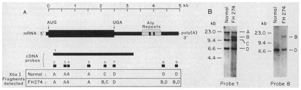
The molecular size of the plasma LDL (low density lipoprotein) receptor synthesized by cultured fibroblasts from a patient with the internalization-defective form of familial hypercholesterolemia (FH 274) was smaller by 10,000 daltons than the size of the normal LDL receptor. The segment of the gene encoding the truncated portion of the FH 274 receptor was cloned into bacteriophage lambda. Comparison of the nucleotide sequences of the normal and FH 274 genes revealed a 5-kilobase deletion, which eliminated the exons encoding the membrane-spanning region and the carboxyl terminal cytoplasmic domain of the receptor. The deletion appeared to be caused by a novel intrastrand recombination between two repetitive sequences of the Alu family that were oriented in opposite directions. The truncated receptors lack membrane-spanning regions and cytoplasmic domains; they are largely secreted into the culture medium, but a small fraction remains adherent to the cell surface. The surface-adherent receptors bind LDL, but they are unable to cluster in coated pits, thus explaining the internalization-defective phenotype.


Figures

Fig. 1
Fig. 1
Analysis of Xba I restriction digests of genomic DNA from FH 274 with probes from different regions of the LDL receptor cDNA. (A) A diagram of the mRNA for the human LDL receptor (9) is shown with AUG and UGA (A, adenine; U, uracil; G, guanine) indicating the beginning and end of the translated region. The hatched areas in the 3′ untranslated region represent Alu repetitive sequences. The sizes and locations of 32P-labeled cDNA probes used to map the LDL receptor gene are shown by the closed bars and are numbered 1 to 9. Probe 1 (double-stranded DNA) was a mixture of a 2.1-kb Eco RI-Sma I fragment and three different 0.9-kb Bam HI-Xho I fragments from p101 (9), which together spanned most of the translated region; the fragments were purified by polyaerylamide gel electrophoresis and electrocution (20) and then labeled with 32P by random hexanucleotide priming (21). Probes 2 to 9 were prepared from M13 subclones of pLDLR-2 (9) as single-stranded, uniformly 32P-labeled DNA, ~100 nucleotides (nt) in length, by the method of Church and Gilbert (22). All probes had a specific radioactivity of at least 5 × 10⁸ cpm/μg. (B) Genomic DNA (5 μg) was isolated from cultured fibroblasts as indicated (20), digested with Xba I (New England Biolabs), subjected to electrophoresis in 1 percent agarose containing buffer A (40 mM tris-acetate, 3 mM disodium EDTA, 20 mM sodium acetate, 18 mM NaCl, pH 8.15), and transferred to nitrocellulose paper (20). The paper was incubated for 16 hours at 42°C with the indicated 32P-labeled cDNA probe (2 × 10⁶ to 4 × 10⁶ cpm/ml) in 50 percent formamide, 1 percent SDS, 5x Denhardt’s solution, 5x (saline, sodium phosphate, and EDTA) (SSPE), and E. coli DNA at 100 μg/ml after preliminary hybridization for 1 hour at 42°C in the same solution without the 32P-labeled probe. After hybridization, the paper was washed in 1 percent SDS plus 2x SSC (saline and sodium citrate) for 15 minutes at 23°C and then in 1 percent SDS plus 0.1 × SSC (probe 1) or in 1 percent SDS plus 0.5 × SSC (probes 2 to 9) for 4 hours at 68°C. Denhardt’s solution, SSPE, and SSC were prepared as described (20). Filters were exposed to x-ray film with an intensifying screen for 24 hours at −70°C. Two representative blot hybridizations (with probes 1 and 8) are shown. The four Xba I restriction fragments are designated A to D along the right side of each blot. Molecular size standards were generated by Hind III cleavage of bacteriophage λ DNA. The Xba I fragments detected by probes 2 to 9 in the normal subject and FH 274 are indicated at the bottom of (A).
Fig. 2
Fig. 2
Blot hybridization of Xba I-cleaved genomic DNA from FH 274 and his parents. Genomic DNA (5 μg) isolated from cultured fibroblasts from the indicated subject was digested with Xba I, subjected to electrophoresis, transferred to nitrocellulose, and hybridized with 32P-labeled probe 1 (see legend to Fig. 1). The four relevant Xba I restriction fragments are designated A to D along the right side of the blot. Molecular size standards were generated by Hind III cleavage of bacteriophage λ DNA.
Fig. 3
Fig. 3
Comparison of restriction maps of the 3′ end of the normal LDL receptor gene and the deletion-bearing gene from FH 274. The scale at the bottom indicates the length of genomic DNA in kilobases. The organization of the normal LDL receptor gene is shown in the diagram at the top. Exons are indicated by solid segments and upper case letters; intervening sequences (IVS) are indicated by open segments and lower case letters. The Alu repetitive sequences in IVSc and exon F are indicated. Restriction enzyme recognition sites used to define the gene deletion in FH 274 are shown. The restriction map of the normal receptor gene was generated from studies of three λ genomic clones (λ 33-2, λ 33-1, λ hl) (10). The map of the gene in FH 274 was generated from λFH 274-10, which contains Xba I fragment B (Fig. 2) (see below). Solid bars above or below the restriction maps denote segments of the normal and mutant genes that were used for DNA sequencing in Figs. 4 and 5. To obtain λFH 274-10, we prepared 570 μg of genomic DNA (20) from fibroblasts of FH 274 and digested it with 1700 units of Xba I. The digested DNA was extracted with a mixture of phenol and chloroform and then chloroform, precipitated with 70 percent ethanol and 86 mM sodium acetate, and dissolved in 200 μl of buffer B (10 mM tris-chloride and 1 mM disodium EDTA at pH 7.5) The DNA (80 μg) was redigested with 100 units of Xba I, and subjected to electrophoresis on a 1 percent “low-gelling-temperature” agarose gel (Bethesda Research Laboratories) containing buffer A (40 V, 72 hours, 4°C). After electrophoresis, ten 2-mm slices of the gel containing DNA fragments, 9 to 23 kb in size, were extracted and concentrated (20), and dissolved in 20 μl of buffer B. One portion (4 μl) of each DNA fraction, 5 μg of Xba I-digested genomic DNA from FH 274, and Hind III-digested λ marker fragments were placed onto individual lanes of a 0.8 percent agarose gel containing buffer A and subjected to electrophoresis (35 V, 16 hours, 23°C). The DNA was transferred to nitrocellulose paper and hybridized with 32P-labeled probe 1 as described in Fig. 1. The resulting autoradiogram identified the fraction that contained the abnormal 13-kb Xba I fragment (fragment B, Fig. 1). The remaining DNA from this fraction (100 ng) was mixed with 500 ng of Xba I-digested arms of λ Charon 35 (23) and incubated (72 hours, 14°C) with 490 units of T4 DNA ligase (New England Biolabs). The ligated material was packaged into λ phage particles in vitro (Amersham) to yield a total of 6.7 × 10³ plaque-forming units. This library was screened with 32P-labeled probe 8 (Fig. 1), which was expected to detect only the abnormal fragment (13 kb) since the corresponding normal fragment (~7 kb) was too small to generate viable recombinant phage. One recombinant clone was identified (λFH 274-10) and isolated after an additional cycle of plaque purification. The 13-kb insert in λFH 274-10 was isolated from purified λ DNA, subcloned into pSP65 (Promega Biotec), and used for restriction endonuclease mapping.
Fig. 4
Fig. 4
Strategy for sequencing and orientation of the relevant Alu repetitive sequences in the normal LDL receptor gene and the corresponding region from the deletion-bearing gene from FH 274. The upper and lower parts of the figure show the relevant regions of the normal and FH 274 genes used for DNA sequencing. These sequences (corresponding to the regions delimited by the solid bars in Fig. 3) were determined from the following clones: IVSc from λ33-1 (10), exon F from the previously reported sequence of pLDLR-2 (9), and the FH 274 deletion joint from λFH 274-10. DNA sequencing was performed by the dideoxy-chain termination method (24). The direction and extent of sequence established in a given experiment are indicated by the long arrows above or below the relevant DNA segments. Those sequences not coinciding with a restriction endonuclease site were determined with synthetic oligonucleotides as primers on M13 subclones (25). The boxed area shows a consensus Alu sequence with the left and right tandem repeats (open and closed, respectively). The nucleotide numbering scheme of the consensus Alu is the same as that described (11); position +1 refers to the C of the Alu I endonuclease recognition site, AGCT. Alu sequences in the normal and FH 274 genes are illustrated according to the same numbering scheme. Orientations of the left and right tandem Alu repeats are indicated by the short arrows. Segments of the normal and FH 274 genes that correspond to each other are connected by the diagonal lines. The exact DNA sequences around nucleotide positions −116 and −7 in the Alu repeats of IVSc and exon F, respectively, and of the deletion joint in FH 274 are shown.
Fig. 5
Fig. 5
Nucleotide sequences across the deletion joint in FH 274 and for the corresponding normal 5′ and 3′ DNA’s. The normal and FH 274 sequence data were obtained as described in Fig. 4 and are numbered according to the scheme used for the consensus Alu sequence (11). The normal 5′ sequence (top line), the sequence across the deletion joint in FH 274 (middle line), and normal 3′ sequence (bottom line) are aligned with only two gaps of one base each. Dots between sequences indicate positions at which the sequences are identical. Horizontal lines above or below the normal sequence indicate positions at which the normal sequence is identical to the consensus Alu sequence (11). The two vertical lines at positions −116 and −7 denote the position of the deletion joint.
Fig. 6
Fig. 6
Potential mechanism for the gene deletion in FH 274. The deletion joint is shown to result from a homologous recombination between the Alu sequences in IVSc and exon F occurring on a single strand of the DNA molecule (steps A–E). The left and right arms of an Alu sequence are indicated by the open and closed bars, respectively. The boxed area at the right shows the actual base pairing that is postulated to occur at step D.
Fig. 7
Fig. 7
Consequences of the 5-kb gene deletion for the structures of the LDL receptor mRNA and truncated protein in FH 274. (A) The location of exons (closed bars) and intervening sequences (IVS, open bars) in the 3′ end of the normal gene and the corresponding segments of the deleted gene in FH 274 are marked. Alu sequences are cross-hatched. The arrows indicate splice sites for IVSc. (B) The translated and untranslated regions (thick and thin closed bars, respectively) of the normal mRNA and corresponding sections of the mutant mRNA are indicated. The location of the 3′ end of the mutant mRNA is unknown (“?”). (C) The structural domains of the normal receptor protein are shown in comparison with those of the mutant receptor protein, which lacks the transmembrane and cytoplasmic domains.
Fig. 8
Fig. 8
Electrophoresis of LDL receptors immunoprecipitated from 35S-labeled fibroblasts from FH 274 and his parents. Fibroblasts were cultured for 6 days and induced for synthesis of LDL receptors by incubation in lipoprotein-deficient serum for 16 hours (4). Cells were pulse-labeled with [35S]methionine (140 μCi/ml) (4) for 2 hours at 37°C, and isotopically labeled LDL receptors were solubilized either immediately (A) or 2 hours after the addition (chase) of unlabeled methionine (B). Cell monolayers were washed once with phosphate-buffered saline at 4°C, and detergent extracts were prepared and incubated with immune complexes (4) containing either monoclonal antibody to LDL receptor (IgG-C7) or a control monoclonal antibody (IgG-2001) (4). The incubation mixtures were layered onto multistep sucrose gradients, and the immunoprecipitates were collected by centrifugation and washed (4). The precipitates were dissolved by heating to 90°C for 5 minutes in buffer containing 10 percent (by volume) glycerol, 0.2M dithiothreitol, 2.3 percent (weight to volume) SDS, and 75 mM tris-chloride at pH 6.8 and subjected to SDS electrophoresis on 7 percent polyacrylamide gels, followed by autoradiography (4). The gels were exposed to x-ray film for 2 days. Apparent molecular sizes of the 35S-labeled proteins were calculated from the position of migration of the following standards as determined by Coomassie blue staining: myosin (200 kD), β-galactosidase (116 kD), phosphorylase b (97 kD), and bovine serum albumin (68 kD). Band z is a protein that appears in control immunoprecipitations and is not related to the LDL receptor; band x is a proteolytic fragment of the receptor.
Fig. 9
Fig. 9
Recovery in the culture medium of newly synthesized LDL receptor from fibroblasts of FH 274 (a) and his mother and father (b). Fibroblasts from the indicated subject were induced for LDL receptor synthesis as described in Fig. 8. Cells were labeled with [35S]methionine (215 μCi/ml for top panel and 275 μCi/ml for bottom panel) for 18 hours at 37°C in medium containing 5 μM unlabeled methionine. The medium (1.1 ml per 60-mm dish) was removed and saved, and each monolayer was washed rapidly twice with a total of 2 ml of phosphate-buffered saline at room temperature. The combined medium and washings from three dishes were pooled and centrifuged at 5000g for 5 minutes at 4°C, and the resulting supernatant was saved (designated medium). Each monolayer was washed an additional four times as above, the washings were discarded, and detergent-extracts from three dishes of each cell strain were prepared as described in Fig. 8. Cell extracts and medium were immunoprecipitated with monoclonal antibody to the receptor (IgG-C7) and analyzed by SDS gel electrophoresis and autoradiography as described in Fig. 8. The gel was exposed to x-ray film for 20 hours.

References

    1. Goldstein JL, Brown MS. In: The Metabolic Basis of Inherited Disease. 5. Stanbury JB, Wyngaarden JB, Fredrickson DS, Goldstein JL, Brown MS, editors. chap 33. McGraw-Hill; New York: 1984. pp. 672–712.
    1. Goldstein JL, Anderson RGW, Brown MS. Nature (London) 1979;279:679. - PubMed
    1. Goldstein JL, Dana SE, Brunschede GY, Brown MS. Proc Natl Acad Sci USA. 1975;72:1092. - PMC - PubMed
    2. Bilheimer DW, Ho YK, Brown MS, Anderson RGW, Goldstein JL. J Clin Invest. 1978;61:678. - PMC - PubMed
    1. Schneider WJ, Beisiegel U, Goldstein JL, Brown MS. J Biol Chem. 1982;257:2664. - PubMed
    2. Tolleshaug H, Goldstein JL, Schneider WJ, Brown MS. Cell. 1982;30:715. - PubMed
    3. Tolleshaug H, Hobgood KK, Brown MS, Goldstein JL. 1983;32:941. ibid. - PubMed
    1. Brown MS, Goldstein JL. Cell. 1976;9:663. - PubMed
    2. Goldstein JL, Brown MS, Stone NJ. 1977;12:629. ibid. - PubMed
    3. Anderson RGW, Goldstein JL, Brown MS. Nature (London) 1977;270:695. - PubMed

Publication types

Associated data

LinkOut - more resources