. 2019 Sep 20;5(9):eaax3009.
doi: 10.1126/sciadv.aax3009. eCollection 2019 Sep.

Multimodal x-ray and electron microscopy of the Allende meteorite

Affiliations

Multimodal x-ray and electron microscopy of the Allende meteorite

Yuan Hung Lo et al. Sci Adv. .

Abstract

Multimodal microscopy that combines complementary nanoscale imaging techniques is critical for extracting comprehensive chemical, structural, and functional information, particularly for heterogeneous samples. X-ray microscopy can achieve high-resolution imaging of bulk materials with chemical, magnetic, electronic, and bond orientation contrast, while electron microscopy provides atomic-scale spatial resolution with quantitative elemental composition. Here, we combine x-ray ptychography and scanning transmission x-ray spectromicroscopy with three-dimensional energy-dispersive spectroscopy and electron tomography to perform structural and chemical mapping of an Allende meteorite particle with 15-nm spatial resolution. We use textural and quantitative elemental information to infer the mineral composition and discuss potential processes that occurred before or after accretion. We anticipate that correlative x-ray and electron microscopy overcome the limitations of individual imaging modalities and open up a route to future multiscale nondestructive microscopies of complex functional materials and biological systems.


Figures

Fig. 1
Fig. 1. Multimodal x-ray and electron nanoscopic spectral imaging scheme.
Allende meteorite grains deposited on a TEM grid were transferred between a Titan 60-300 electron microscope and the COSMIC soft x-ray beamline for tomographic, ptychographic, and spectromicroscopic imaging. COSMIC’s TEM-compatible sample holder enabled the same meteorite grain to be imaged using both imaging modalities to extract multidimensional datasets, providing chemical, structural, and functional insights with high spatial resolution.
Fig. 2
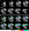
Fig. 2. HAADF and EDS GENFIRE tomography reconstructions.
Representative 14-nm-thick layers in the reconstructed 3D HAADF (A) and EDS (B) volumes of the Allende meteorite grain. The red arrow points to melt pockets, and the green arrow points to shock veins that were embedded, which suggest that the sample had at some point experienced impact-induced heating, cracking, and melting. The traces of aluminum and chromium in the veins that are visible in the EDS reconstructions reveal that the veins were filled with metallic recrystallization. a.u., arbitrary units.
Fig. 3
Fig. 3. X-ray ptychography and STXM absorption spectromicroscopy.
(A to D) Localization of major elements in the meteorite revealed by dividing pre-edge and on-edge ptychography images at the absorption edges for Al, Fe, Mg, and Ni. The absorption quotient maps, displayed in logarithmic scale, show the presence of Fe in the shock veins of the silicate that is barely observable in EDS images (red arrows). (E and F) Scattering quotient (fq) maps derived from ptychographic Mg pre-edge and Al pre-edge images, respectively. This region of interest is a zoomed-in view from the dashed red rectangle shown in (B). (G and H) Ni-Fe ratio maps from Mg pre-edge and Al pre-edge scattering quotient maps, respectively. These ratio maps are converted using the SQUARREL method, given a fixed amount of sulfur. The color bar indicates the Ni-Fe ratio and 100% implies a pure nickel sulfide region. (I to L) Absorption spectra generated from STXM energy scans across the four absorption edges, revealing unique spectral fingerprints for each respective element and also showing pronounced spectral differences in the different iron-containing regions. Relative peak intensities between Fe L3a and L3b also reveal the presence of predominant Fe2+ species. Colors of the solid lines match colors of mineral regions in Fig. 2B.
Fig. 4
Fig. 4. Possible grain composition based on EDS quantification of elemental abundances.
Ternary plots of major elements as quantified by the Cliff-Lorimer method for three different mineral types in the meteoric grain. Quantitative compositional information narrows down the possible mineral types and suggests that the sulfide is similar to pentlandite (A), the silicate is similar to ferrosilite (B), and the oxide is a chromium spinel or chromite (C). wt %, weight %.

References

    1. Miao J., Ishikawa T., Robinson I. K., Murnane M., Beyond crystallography: Diffractive imaging using coherent x-ray light sources. Science 348, 530–535 (2015). - PubMed
    1. Pfeiffer F., X-ray ptychography. Nat. Photonics 12, 9–17 (2018).
    1. Falcone R., Jacobsen C., Kirz J., Marchesini S., Shapiro D., Spence J., New directions in X-ray microscopy. Contemp. Phys. 52, 293–318 (2011).
    1. Hitchcock A. P., Soft X-ray spectromicroscopy and ptychography. J. Electron Spectrosc. Relat. Phenom. 200, 49–63 (2015).
    1. Miao J., Charalambous P., Kirz J., Sayre D., Extending the methodology of X-ray crystallography to allow imaging of micrometre-sized non-crystalline specimens. Nature 400, 342–344 (1999).

Publication types