Review
. 2020 Apr 1;41(2):371-403.
doi: 10.1210/endrev/bnz004.

Advances in the Diagnosis and Management of Well-Differentiated Neuroendocrine Neoplasms

Affiliations
Review

Advances in the Diagnosis and Management of Well-Differentiated Neuroendocrine Neoplasms

Johannes Hofland et al. Endocr Rev. .

Abstract

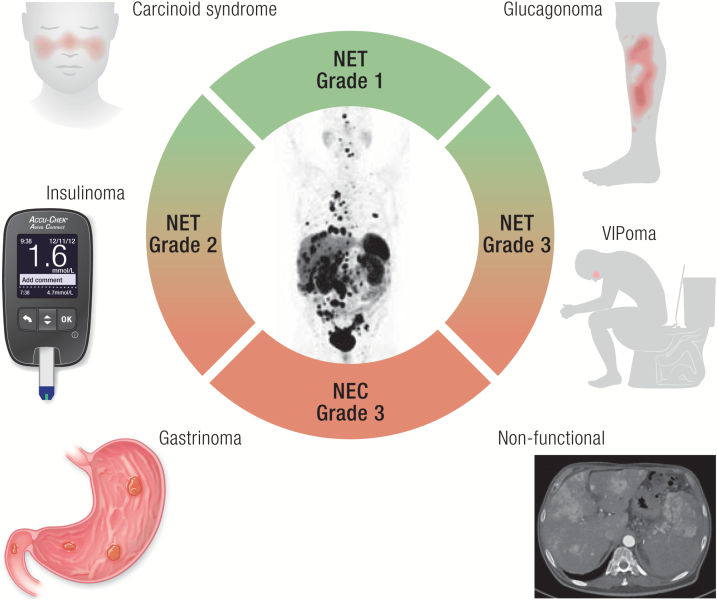
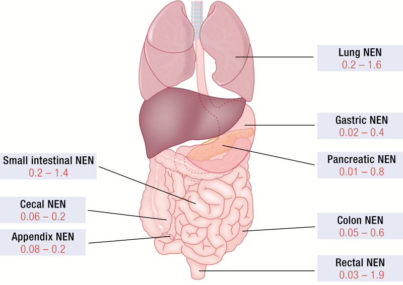
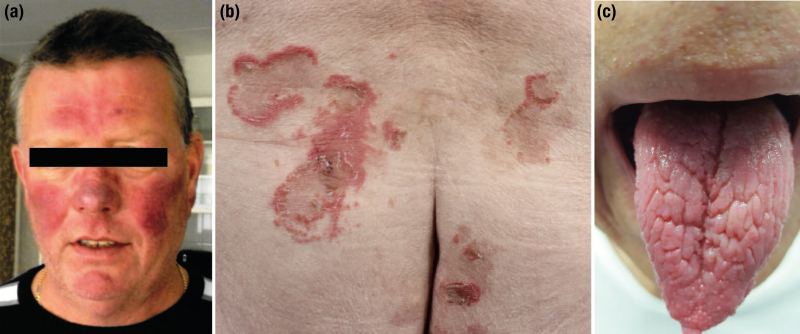
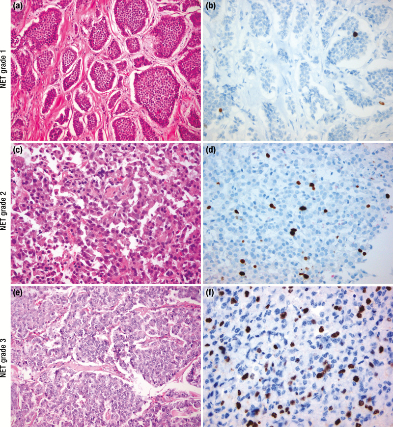
Neuroendocrine neoplasms constitute a diverse group of tumors that derive from the sensory and secretory neuroendocrine cells and predominantly arise within the pulmonary and gastrointestinal tracts. The majority of these neoplasms have a well-differentiated grade and are termed neuroendocrine tumors (NETs). This subgroup is characterized by limited proliferation and patients affected by these tumors carry a good to moderate prognosis. A substantial subset of patients presenting with a NET suffer from the consequences of endocrine syndromes as a result of the excessive secretion of amines or peptide hormones, which can impair their quality of life and prognosis. Over the past 15 years, critical developments in tumor grading, diagnostic biomarkers, radionuclide imaging, randomized controlled drug trials, evidence-based guidelines, and superior prognostic outcomes have substantially altered the field of NET care. Here, we review the relevant advances to clinical practice that have significantly upgraded our approach to NET patients, both in diagnostic and in therapeutic options.


Figures

Graphical Abstract
Graphical Abstract
Figure 1.
Figure 1.
Neuroendocrine neoplasms (NEN) locations and incidence rates. The most common primary NEN sites of the pulmonary and gastroenteropancreatic systems are depicted. Incidence rates were collected from Fraenkel et al. (9). and Dasari et al. (8). and are shown in red as the incident number of cases per 100 000 per year.
Figure 2.
Figure 2.
Clinical signs of hormonal excess in neuroendocrine neoplasms (NENs). (A) Facial flushing in the context of carcinoid syndrome in a patient with a metastasized midgut neuroendocrine tumor. (B) Necrolytic migratory erythema at the sacral region and (C) glossitis in a patient with a metastasized glucagonoma.
Figure 3.
Figure 3.
Histopathology of neuroendocrine neoplasms (NENs). Hematoxylin and eosin (H&E, A) and Ki67 (B) staining of a grade 1 NET showing nests of neuroendocrine cells with oval nuclei, “salt and paper” chromatin and moderate eosinophilic cytoplasm. The nests are separated by a fibrous stroma. Nuclear staining of Ki67 is only visible in a few neoplastic cells (Ki67 <3%). (C) Histology of a grade 2 neuroendocrine tumor (NET) reveals a homogeneous population of neuroendocrine cells with slight atypia, round to oval nuclei, dense chromatin and moderate eosinophilic/ amphophilic cytoplasm. (D) Ki67 staining in the same tumor revealed 5% positivity in a hotspot. (E) A grade 3 NET displays a well-differentiated histology of neuroendocrine cells with vesicular nuclei without nucleoli and moderate amphophilic cytoplasm arranged in a nested pattern, whereas the Ki67 proliferation index is above 20% (F). H&E and Ki67 images are amplified ×200 and ×400, respectively.
Figure 4.
Figure 4.
Imaging procedures used in neuroendocrine neoplasm (NEN) diagnostics. (A) Axial T2-weighted magnetic resonance imaging (MRI) showing metastatic deposits in both hepatic lobes from a pancreatic Grade 2 NET. (B) Axial T1 diffusion MRI image of the same patient showing further lesions not detected with the previous MRI sequence. (C) Computed tomography (CT) of the abdomen demonstrating a desmoplastic reaction (white arrow) in the mesentery of a patient with a Grade 1 small bowel NET. (D) Fibrotic strands radiating from a central mesenteric metastatic mass in a patient with multiple small bowel NETs. There is thickening of the bowel wall and fluid retention due to venous ischemia in this patient, causing postprandial abdominal pain. (E) MRI T2-weighted image with fat saturation demonstrating an oval shaped high signal bone lesion from a Grade 2 small bowel NET at the level of Th11 (white arrow). (F) Polypoid lesion arising from the body of the stomach detected by endoscopic ultrasonography infiltrating the mucosa and submucosa. (G) Positive right hepatic lobe 18F-FDG PET uptake (white arrow) in a patient with a small bowel Grade 2 NET. In the same patient positive 68Gallium-DOTATOC positron emission tomography (PET) in the same area of 18F-FDG PET uptake (thick white arrow) and additional uptake in different areas of the left hepatic lobe (thin white arrow). (H) Positive uptake in multiple hepatic areas in a patient with a Grade 2 pancreatic NET following a 68Gallium-DOTATOC PET. Negative 18F-FDG PET in tumor lesions within the same patient.
Figure 5.
Figure 5.
Diagnostic algorithm. Histology should be obtained from tumors suspected of NEN to confirm the diagnosis of a neuroendocrine origin. Morphological examination will subsequently divide neoplasms into well-differentiated tumors or poorly differentiated carcinomas. Uncertain cases can be categorized through the use of genetic analysis or p53 staining. Within the NETs mitotic and Ki-67 indices will classify the tumor into grade 1 to 3. Further prognostic and therapeutic information can be obtained by performing 68Ga-labelled somatostatin receptor imaging and for higher grade or clinically aggressive tumors an 18F-FDG PET. FDG, fluorodeoxyglucose NEN, neuroendocrine neoplasm; WD, well-differentiated; PD, poorly differentiated; NET, neuroendocrine tumor; NEC, neuroendocrine carcinoma; SUV, standardized uptake value; PET, positron emission tomography; Pan, pancreas; GI, gastrointestinal.
Figure 6.
Figure 6.
Therapeutic targets for neuroendocrine neoplasms (NENs). Overview of the different therapeutic modalities for proliferative control in NENs and their respective targets within the NEN cell.

References

    1. Perren A, Couvelard A, Scoazec JY, et al. ; Antibes Consensus Conference participants . ENETS consensus guidelines for the standards of care in neuroendocrine tumors: pathology: diagnosis and prognostic stratification. Neuroendocrinology. 2017;105(3):196–200. - PubMed
    1. Inzani F, Petrone G, Rindi G. The New World Health Organization classification for pancreatic neuroendocrine neoplasia. Endocrinol Metab Clin North Am. 2018;47(3):463–470. - PubMed
    1. Kaltsas GA, Besser GM, Grossman AB. The diagnosis and medical management of advanced neuroendocrine tumors. Endocr Rev. 2004;25(3):458–511. - PubMed
    1. Asa SL, Casar-Borota O, Chanson P, et al. ; attendees of 14th Meeting of the International Pituitary Pathology Club, Annecy, France, November 2016. From pituitary adenoma to pituitary neuroendocrine tumor (PitNET): an International Pituitary Pathology Club proposal. Endocr Relat Cancer. 2017;24(4):C5–C8. - PubMed
    1. Schadendorf D, Lebbé C, Zur Hausen A, et al. Merkel cell carcinoma: epidemiology, prognosis, therapy and unmet medical needs. Eur J Cancer. 2017;71:53–69. - PubMed

Publication types

MeSH terms