Review
. 2019 Oct 7;48(39):14547-14565.
doi: 10.1039/c9dt03039e.

A brief overview of metal complexes as nuclear imaging agents

Affiliations
Review

A brief overview of metal complexes as nuclear imaging agents

Douglas S MacPherson et al. Dalton Trans. .

Abstract

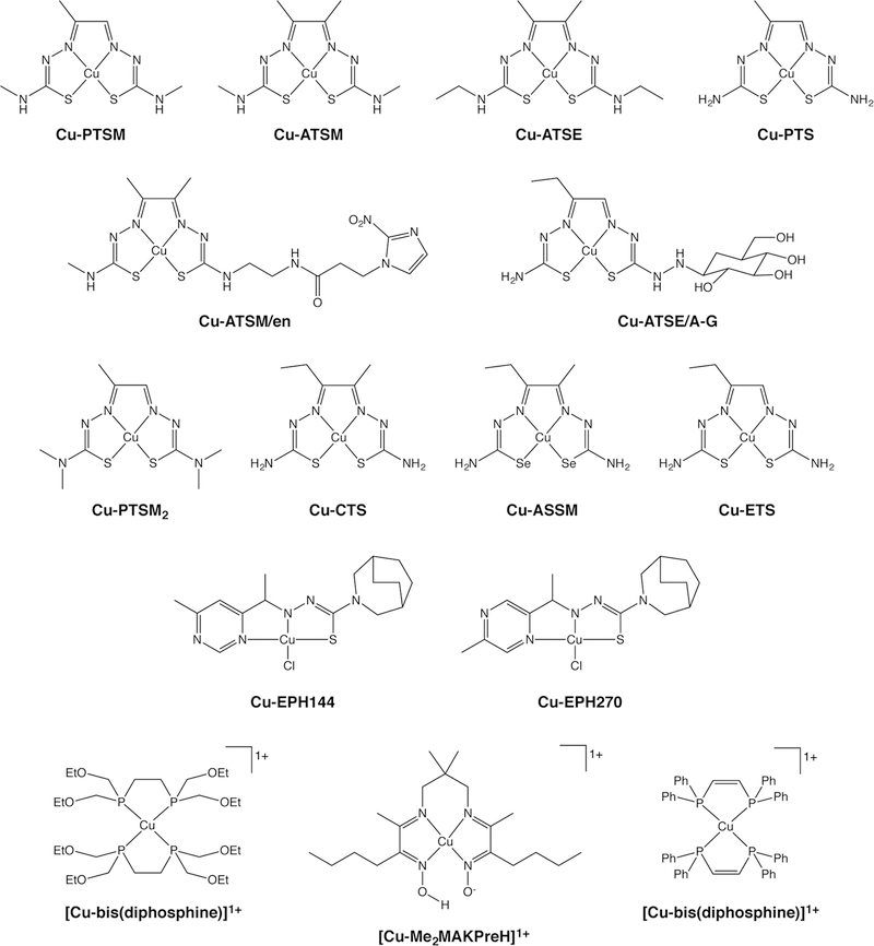
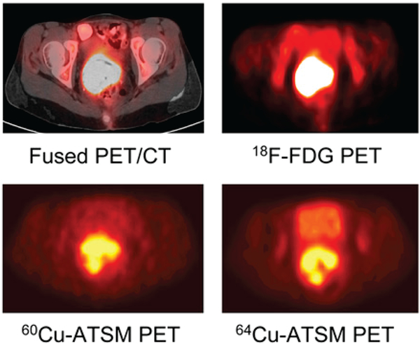
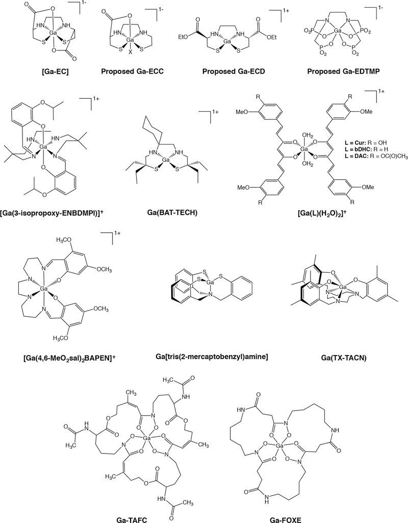
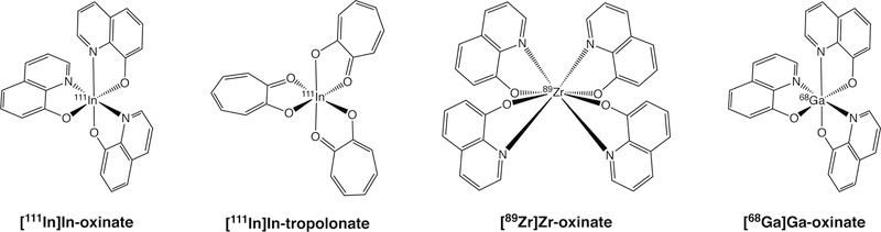
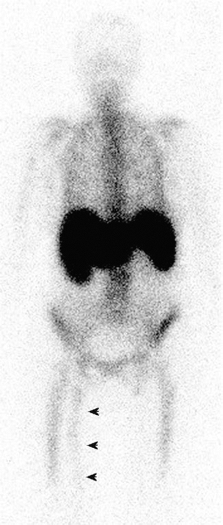
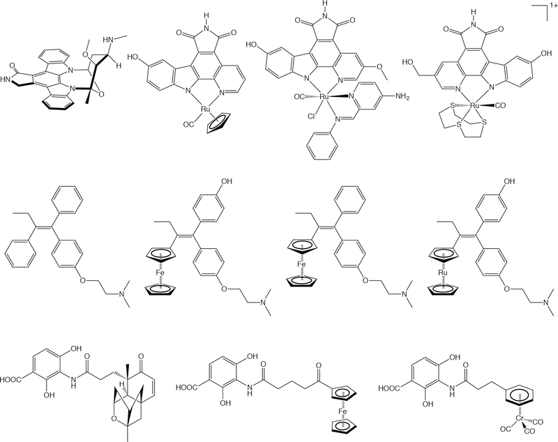
Metallic radionuclides have been instrumental in the field of nuclear imaging for over half a century. While recent years have played witness to a dramatic rise in the use of radiometals as labels for chelator-bearing biomolecules, imaging agents based solely on coordination compounds of radiometals have long played a critical role in the discipline as well. In this work, we seek to provide a brief overview of metal complex-based radiopharmaceuticals for positron emission tomography (PET) and single photon emission computed tomography (SPECT). More specifically, we have focused on imaging agents in which the metal complex itself rather than a pendant biomolecule or targeting moiety is responsible for the in vivo behavior of the tracer. This family of compounds contains metal complexes based on an array of different nuclides as well as probes that have been used for the imaging of a variety of pathologies, including infection, inflammation, cancer, and heart disease. Indeed, two of the defining traits of transition metal complexes-modularity and redox chemistry-have both been creatively leveraged in the development of imaging agents. In light of our audience, particular attention is paid to structure and mechanism, though clinical data is addressed as well. Ultimately, it is our hope that this review will not only educate readers about some of the seminal work performed in this space over the last 30 years but also spur renewed interest in the creation of radiopharmaceuticals based on small metal complexes.


Figures

Figure 1.
Figure 1.
Selected [99mTc]Tc(I) and [99mTc]Tc(III) complexes
Figure 2.
Figure 2.
Planar [99mTc]Tc-MIBI scintigraphy (A and B), CT (C), and SPECT/CT (D) of a patient with primary hyperparathyroidism, showing focal tracer uptake in the lower part of the right lobe of the thyroid gland. Reprinted from Keidar, et al. Preoperative [99mTc]Tc-MIBI SPECT/CT interpretation criteria for localization of parathyroid adenomas — correlation with surgical findings. Molecular Imaging and Biology 2017, 19, 265–270. Copyright by the World Molecular Imaging Society.
Figure 3.
Figure 3.
Selected [99mTc]Tc(IV) and [99mTc]Tc(V) complexes
Figure 4.
Figure 4.
Planar, whole-body [99mTc]TcN-DBODC5 scintigraphy (left to right: collected 5, 30, 60, and 240 min post-injection) of a healthy under (A) rest and (B) stress conditions. Adapted and reprinted from Cittanti et al. Whole-body biodistribution and radiation dosimetry of the new cardiac tracer 99mTcN-DBODC. Journal of Nuclear Medicine 2008, 49, 1299–1304. Copyright by the Society of Nuclear Medicine and Molecular Imaging.
Figure 5.
Figure 5.
Selected Cu(II) complexes
Figure 6.
Figure 6.
Transaxial [18F]FDG-PET/CT, [18F]FDG PET, [60Cu]Cu-ATSM PET, and [64Cu]Cu-ATSM images of a patient with cancer of the uterine cervix. 18F-FDG = [18F]fluorodeoxyglucose = 2-deoxy-2-[18F]fluoroglucose. Adapted and reprinted from Lewis et al. An imaging comparison of [64Cu]Cu-ATSM and [60Cu]Cu-ATSM in cancer of the uterine cervix. Journal of Nuclear Medicine 2008, 49 (7), 1177–1182. Copyright by the Society of Nuclear Medicine and Molecular Imaging.
Figure 7.
Figure 7.
Selected Ga(III) complexes
Figure 8.
Figure 8.
Selected [111In]In(III), [89Zr]Zr(IV), and [68Ga]Ga(III) complexes
Figure 9.
Figure 9.
Planar scintigraphy using [111In]In-oxine-labeled leukocytes revealing an area of increased uptake in an infected prosthetic vascular graft in the right thigh (black arrows). Adapted and reprinted from Love et al. Radionuclide imaging of infection. Journal of Nuclear Medicine Technology 2004, 32, 47–57. Copyright by the Society of Nuclear Medicine and Molecular Imaging.
Figure 10.
Figure 10.
Selected medicinal inorganic and organometallic compounds. Top: Ru-based kinase inhibitors inspired by the structure of staurosporine (far left). Middle: Fe- and Ru-based selective estrogen receptor modulators inspired by the structure of tamoxifen (far left). Bottom: Fe- and Cr-based antibiotics based on the structure of platensimycin (far left).

References

    1. Wadas TJ, Wong EH, Weisman GR and Anderson CJ, Chem. Rev, 2010, 110, 2858–2902. - PMC - PubMed
    1. Zeglis BM and Lewis JS, Dalton Trans, 2011, 40, 6168–6195. - PMC - PubMed
    1. Liu S and Chakraborty S, Dalton Trans, 2011, 40, 6077–6086. - PubMed
    1. Blower PJ, Dalton Trans, 2015, 44, 4819–4844. - PMC - PubMed
    1. Paterson BM and Donnelly PS, Chem. Soc. Rev, 2011, 40, 3005–3018. - PubMed

MeSH terms

LinkOut - more resources