. 2019 Dec 1;21(6):526-534.
doi: 10.1001/jamafacial.2019.0851.

Molecular Characterization of Lipoaspirates Used in Regenerative Head and Neck Surgery

Affiliations

Molecular Characterization of Lipoaspirates Used in Regenerative Head and Neck Surgery

Kariem Sharaf et al. JAMA Facial Plast Surg. .

Abstract

Importance: Adipose-derived mesenchymal stem cells (ASCs) have been used commonly in regenerative medicine and increasingly for head and neck surgical procedures. Lipoaspiration with centrifugation is purported to be a mild method for the extraction of ASCs used for autologous transplants to restore tissue defects or induce wound healing. The content of ASCs, their paracrine potential, and cellular potential in wound healing have not been explored for this method to our knowledge.

Objective: To evaluate the characteristics of lipoaspirates used in reconstructive head and neck surgical procedures with respect to wound healing.

Design, setting, and participants: This case series study included 15 patients who received autologous fat injections in the head and neck during surgical procedures at a tertiary referral center. The study was performed from October 2017 to November 2018, and data were analyzed from October 2017 to February 2019.

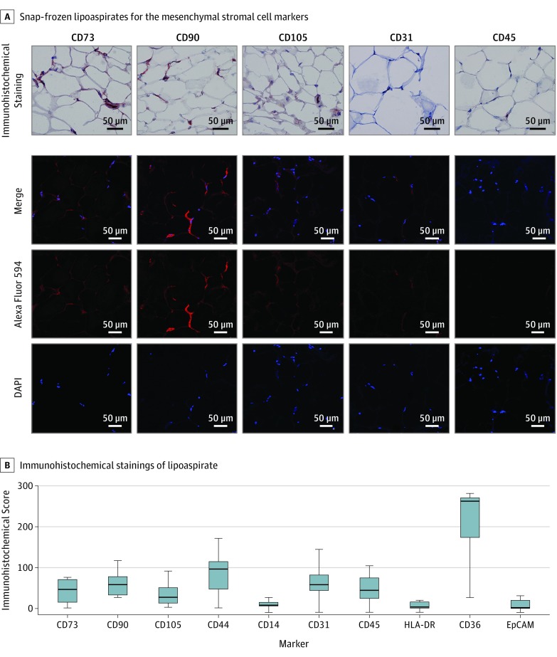
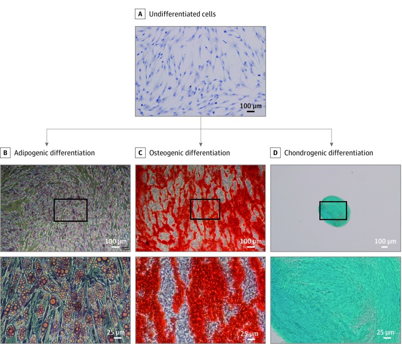
Main outcomes and measures: Excessive material of lipoaspirates from subcutaneous abdominal fatty tissue was examined. Cellular composition was analyzed using immunohistochemistry (IHC) and flow cytometry, and functionality was assessed through adipose, osteous, and chondral differentiation in vitro. Supernatants were tested for paracrine ASC functions in fibroblast wound-healing assays. Enzyme-linked immunosorbent assay measurement of tumor necrosis factor (TNF), vascular endothelial growth factor (VEGF), stromal-derived factor 1α (SDF-1α), and transforming growth factor β3 (TGF-β3) was performed.

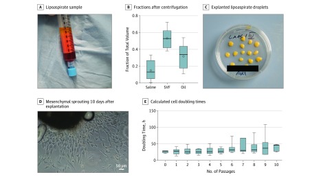
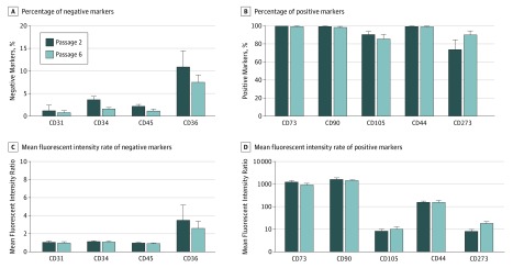
Results: Among the 15 study patients (8 [53.3%] male; mean [SD] age at the time of surgery, 63.0 [2.8] years), the stromal vascular fraction (mean [SE], 53.3% [4.2%]) represented the largest fraction within the native lipoaspirates. The cultivated cells were positive for CD73 (mean [SE], 99.90% [0.07%]), CD90 (99.40% [0.32%]), and CD105 (88.54% [2.74%]); negative for CD34 (2.70% [0.45%]) and CD45 (1.74% [0.28%]) in flow cytometry; and negative for CD14 (10.56 [2.81] per 300 IHC score) and HLA-DR (6.89 [2.97] per 300 IHC score) in IHC staining; they differentiated into osteoblasts, adipocytes, and chondrocytes. The cultivated cells showed high expression of CD44 (mean [SE], 99.78% [0.08%]) and CD273 (82.56% [5.83%]). The supernatants were negative for TNF (not detectable) and SDF-1α (not detectable) and were positive for VEGF (mean [SE], 526.74 [149.84] pg/mL for explant supernatants; 528.26 [131.79] pg/106 per day for cell culture supernatants) and TGF-β3 (mean [SE], 22.79 [3.49] pg/mL for explant supernatants; 7.97 [3.15] pg/106 per day for cell culture supernatants). Compared with control (25% or 50% mesenchymal stem cell medium), fibroblasts treated with ASC supernatant healed the scratch-induced wound faster (mean [SE]: control, 1.000 [0.160]; explant supernatant, 1.369 [0.070]; and passage 6 supernatant, 1.492 [0.094]).

Conclusions and relevance: The cells fulfilled the international accepted criteria for mesenchymal stem cells. The lipoaspirates contained ASCs that had the potential to multidifferentiate with proliferative and immune-modulating properties. The cytokine profile of the isolated ASCs had wound healing-promoting features. Lipoaspirates may have a regenerative potential and an application in head and neck surgery.

Level of evidence: NA.


Conflict of interest statement

Conflict of Interest Disclosures: Dr Haubner reported receiving personal fees from Spiggle & Theis outside the submitted work. No other disclosures were reported.

Figures

Figure 1.
Figure 1.. Explant Culture and Isolation
A, Lipoaspirate from independent samples (n = 8) after centrifugation, with clear separation between oil, stromal vascular fraction, and saline fraction. B, Graph depicts the mean (SE) volume distribution of 8 lipoaspirate fractions. Box plots show the 25th to 75th percentiles, with whiskers indicating minimum and maximum values, horizontal lines in the boxes indicating medians, and circles indicating means. C, Representative tissue culture dish with explanted lipoaspirate droplets. D, Cells with a predominantly mesenchymal phenotype sprouting from 1 representative explanted lipoaspirate 10 days after explantation (original magnification ×100). E, Mean (SE) doubling times calculated from cell counting with viability staining from explant cultures (n = 6 independent samples). Box plots show the 25th to 75th percentiles, with whiskers indicating minimum and maximum values and horizontal lines in the boxes indicating medians. Oil indicates oil from dissolving lipocytes due to centrifugation; and SVF, stromal-vascular fraction.
Figure 2.
Figure 2.. Immunohistochemical Staining of Lipoaspirates
A, Immunohistochemical (brown; top) and hemalaun (blue; top) staining and confocal imaging of snap frozen lipoaspirate samples for the mesenchymal stromal cell markers CD73, CD90, and CD105; endothelial cell marker CD31; and leukocyte common antigen CD45 (original magnification ×200). The 4′,6-diamidino-2-phenylindole (DAPI) signal is shown in blue and Alexa Flour 594 signal in red. B, Immunohistochemical scores for stained lipoaspirates (n = 9) for the indicated markers. Box plots show the 25th to 75th percentiles, with whiskers indicating minimum and maximum values and horizontal lines in the boxes indicating medians. EpCAM indicates epithelial cell adhesion molecule.
Figure 3.
Figure 3.. Flow Cytometry of Adipose Tissue–Derived Isolated Explant Cells for 6 Independent Samples
A and B, Percentages of positive populations of passages 2 and 6 adipose-derived mesenchymal stem cells (ASCs) were compared for the expression of the indicated markers. C and D, Mean (SE) fluorescence intensity ratios of passages 2 and 6 ASCs were compared for the expression of the indicated markers. aP ≤ .05.
Figure 4.
Figure 4.. Multilineage Differentiation Potential In Vitro of ASCs Into Adipocytes, Osteocytes, and Chondrocytes
A, Undifferentiated passage 2 adipose-derived mesenchymal stem cells (ASCs) grown on glass slides and stained with Mayer hematoxylin counterstain. One representative image from 3 samples is shown (original magnification ×100). B, Lipid vesicle accumulation visualized by Oil Red O staining in adipogenic differentiated ASCs after 2 weeks of incubation with adipogenic differentiation medium (original magnification ×50 and ×200). C, Alizarin S staining of extracellular calcium deposits in osteogenic differentiated ASCs after 2 weeks of incubation with osteogenic differentiation medium (original magnification ×50 and ×200). D, Alcian blue staining of cartilage extracellular matrix in ASCs cultured as 3-dimensional spheroids after 4 weeks of incubation with chondrogenic differentiation medium (original magnification ×40 and ×100). Representative images of 6 independent experiments are shown. The area of higher magnification is indicated with the black boxes in the upper images of lower magnification.
Figure 5.
Figure 5.. Exocrine Functions of Explant Culture and Isolated Cells
A, Representative examples of wound areas at 0 and 24 hours for control cells and after incubation with explant supernatants or cell culture supernatants collected at passage 6; 25% and 50% supernatant concentrations were used. Representative images of 3 experiments are shown (original magnification ×50). B, Cell migration date extrapolated from wound-healing assay was compared. C, Enzyme-linked immunosorbent assay results for vascular endothelial growth factor (VEGF), transforming growth factor β3 (TGF-β3), stromal-derived factor 1α (SDF-1α), and tumor necrosis factor (TNF) in explant and ASC culture supernatants collected in passages 1 and 6. aP ≤ .05, by 1-way analysis of variance and the Dunnet multiple comparison test (n = 3). bP ≤ .01, by 1-way analysis of variance and the Dunnet multiple comparison test (n = 3). cResults below the assay range.

Similar articles

Cited by

References

    1. Bui P, Lepage C. Benefits of volumetric to facial rejuvenation. Part 1: fat grafting [in French]. Ann Chir Plast Esthet. 2017;62(5):532-549. doi:10.1016/j.anplas.2017.07.004 - DOI - PubMed
    1. Delay E, Savu T, Atanasiu M. Lipomodeling in breast reconstruction [in French]. Ann Chir Plast Esthet. 2018;63(5-6):505-515. doi:10.1016/j.anplas.2018.06.012 - DOI - PubMed
    1. Krastev TK, Beugels J, Hommes J, Piatkowski A, Mathijssen I, van der Hulst R. Efficacy and safety of autologous fat transfer in facial reconstructive surgery: a systematic review and meta-analysis. JAMA Facial Plast Surg. 2018;20(5):351-360. doi:10.1001/jamafacial.2018.0102 - DOI - PMC - PubMed
    1. Bashir MM, Sohail M, Bashir A, et al. . Outcome of conventional adipose tissue grafting for contour deformities of face and role of ex vivo expanded adipose tissue-derived stem cells in treatment of such deformities. J Craniofac Surg. 2018;29(5):1143-1147. doi:10.1097/SCS.0000000000004367 - DOI - PubMed
    1. Hespe GE, Albornoz CR, Mehrara BJ, Kraus D, Matros E. Pharyngocutaneous fistula closure using autologous fat grafting. Eplasty. 2013;13:e23. - PMC - PubMed