. 2019 Oct 22;13(10):12148-12161.
doi: 10.1021/acsnano.9b06691. Epub 2019 Sep 30.

Positron Emission Tomography-Guided Photodynamic Therapy with Biodegradable Mesoporous Silica Nanoparticles for Personalized Cancer Immunotherapy

Affiliations

Positron Emission Tomography-Guided Photodynamic Therapy with Biodegradable Mesoporous Silica Nanoparticles for Personalized Cancer Immunotherapy

Cheng Xu et al. ACS Nano. .

Abstract

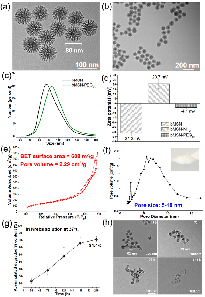
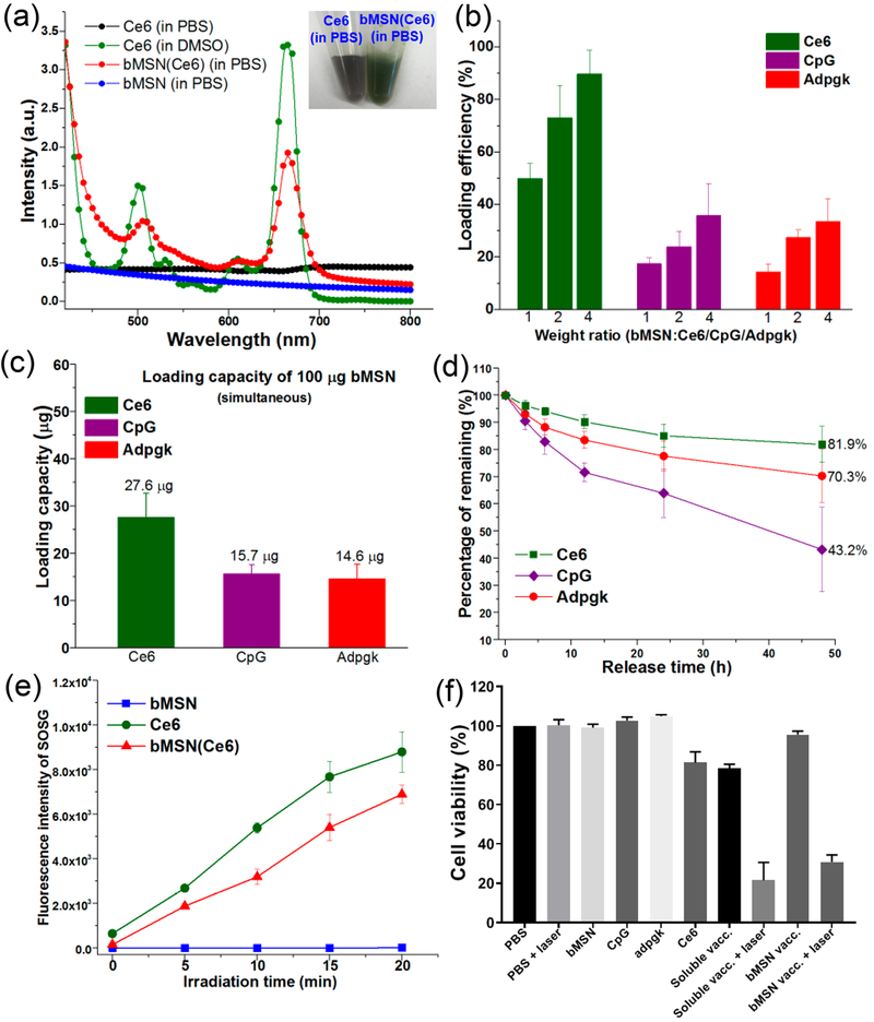
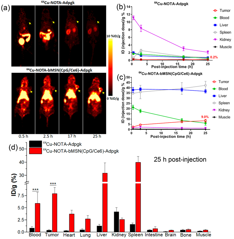
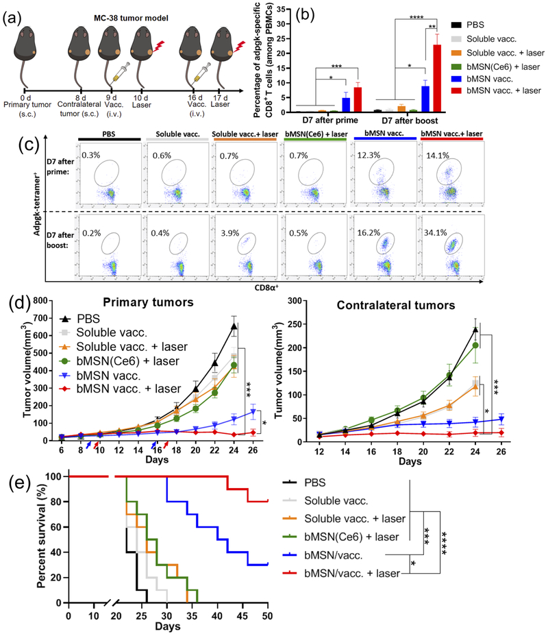
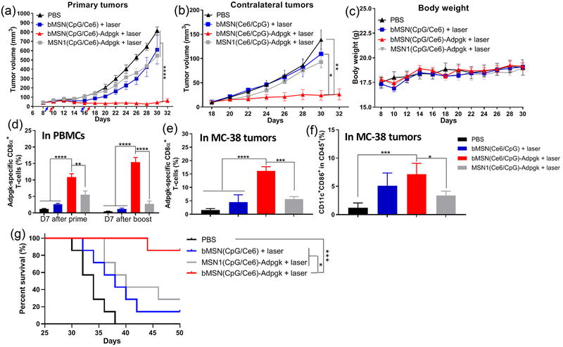
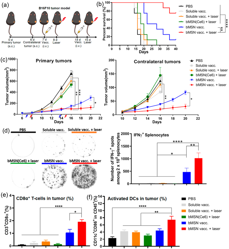
Photodynamic therapy (PDT) is an effective, noninvasive therapeutic modality against local tumors that are accessible to the source of light. However, it remains challenging to apply PDT for the treatment of disseminated, metastatic cancer. On the other hand, cancer immunotherapy offers a promising approach for generating systemic antitumor immune responses against disseminated cancer. Here we report a multifunctional nanomaterial system for the combination of PDT and personalized cancer immunotherapy and demonstrate their potency against local as well as disseminated tumors. Specifically, we have synthesized uniform and biodegradable mesoporous silica nanoparticles (bMSN) with an average size of ∼80 nm and large pore size of 5-10 nm for theranostic positron emission tomography (PET)-guided PDT and neoantigen-based cancer vaccination. Multiple neoantigen peptides, CpG oligodeoxynucleotide adjuvant, and photosensitizer chlorin e6 were coloaded into a bMSN nanoplatform, and PET imaging revealed effective accumulation of bMSN in tumors (up to 9.0% ID/g) after intravenous administration. Subsequent PDT with laser irradiation recruited dendritic cells to PDT-treated tumor sites and elicited neoantigen-specific, tumor-infiltrating cytotoxic T-cell lymphocytes. Using multiple murine models of bilateral tumors, we demonstrate strong antitumor efficacy of PDT-immunotherapy against locally treated tumors as well as distant, untreated tumors. Our findings suggest that the bMSN is a promising platform for combining imaging and PDT-enhanced personalized immunotherapy for the treatment of advanced cancer.

Keywords: cancer immunotherapy; mesoporous silica nanoparticles; neoantigen; photodynamic therapy; positron emission tomography; vaccine.


Conflict of interest statement

The authors declare no competing financial interest.

Figures

Figure 1.
Figure 1.
Schematic illustration of fabrication of bMSN(CpG/Ce6)-neoantigen and mechanism of bMSN(CpG/Ce6)-neoantigen nanovaccines for PDT-enhanced cancer immunotherapy. We synthesized bMSN using the heterogeneous oil–water biphasic reaction system. CpG and Ce6 were loaded into the mesopores of bMSNs through electrostatic and hydrophobic interactions, respectively. After surface PEGylation of bMSNs with PDP-PEG5k-NHS, neoantigen peptides were conjugated to bMSNs via formation of disulfide bonds. Laser irradiation (660 nm) was applied to generate cytotoxic ROS and eliminate tumor cells, while triggering local immune activation for antitumor immunity.
Figure 2.
Figure 2.
Morphology, structure, and characterization of bMSNs. (a and b) TEM images of bMSNs; (c) hydrodynamic size analysis of bMSNs (black line) and bMSN-PEG (green line) by DLS; (d) surface zeta potential of bMSNs, bMSN-NH2, and bMSN-PEG; nitrogen adsorption and desorption isotherms (e) and pore size distributions (f) of bMSNs; (g) in vitro biodegradation profile of bMSNs in simulated body fluid (Krebs–Henseleit solution) at 37 °C for 9 days. At indicated time points, TEM images of (h) were obtained.
Figure 3.
Figure 3.
(a) UV–vis absorption spectra of Ce6 (in PBS), Ce6 (in DMSO), bMSN(Ce6) (in PBS), and bMSN (in PBS). Inset: Photographs of Ce6 (in PBS) and bMSN(Ce6) (in PBS) with a Ce6 concentration of 0.2 mg/mL. (b) Loading efficiency of CpG, Ce6, and Adpgk peptide by bMSNs in various weight ratios. (c) Simultaneous loading capacity of CpG, Ce6, and Adpgk peptide by 100 μg of bMSNs in PBS. (d) Release profile of bMSN(CpG/Ce6)-Adpgk in PBS at 37 °C. (e) Singlet oxygen production by bMSNs, free Ce6, and bMSN(Ce6) after 660 nm laser irradiation (25 mW/cm2) as measured by the changes in the fluorescence intensity of SOSG (Singlet Oxygen Sensor Green). (f) In vitro PDT assay after incubating MC-38 tumor cells with bMSNs (0.5 mg/mL), Ce6 (0.5 μg/mL), CpG (1.0 μg/mL), Adpgk (10 μg/mL), soluble vaccine (CpG (1.0 μg/mL), Ce6 (1.0 μg/mL), Adpgk (10 μg/mL)) with or without laser irradiation (660 nm, 10 mW/cm2, 2 min), and bMSN vaccine (bMSN(CpG/Ce6)-Adpgk, same Ce6, CpG, Adpgk concentration with soluble vaccine) with or without laser irradiation (660 nm, 10 mW/cm2, 2 min).
Figure 4.
Figure 4.
(a) Serial PET images of MC-38 tumor-bearing mice at various time points postinjection of 64Cu-NOTA-Adpgk or 64Cu-NOTA-bMSN(CpG/Ce6)-Adpgk. Tumors are indicated by yellow arrowheads. Time–radioactivity curves of MC-38 tumor, blood, liver, spleen, kidney, and muscle after i.v. injection of 64Cu-NOTA-Adpgk (b) and 64Cu-NOTA-bMSN(CpG/Ce6)-Adpgk (c). (d) Biodistribution studies in MC-38 tumor-bearing mice at 25 h postinjection of 64Cu-NOTA-Adpgk and 64Cu-NOTA-bMSN(CpG/Ce6)-Adpgk.
Figure 5.
Figure 5.
Antitumor therapy study in MC-38 tumor-bearing mice. (a) C57BL/6 mice were randomly divided into the following six treatment groups: (1) PBS control; (2) soluble vaccine (CpG, Ce6, and Adpgk peptide); (3) soluble vaccine with laser irradiation; (4) bMSN(Ce6) with laser irradiation; (5) bMSN vaccine (bMSN(CpG/Ce6)-Adpgk); and (6) bMSN vaccine with laser irradiation. Laser irradiation (660 nm, 50 mW/cm2 for 15 min) was conducted over the tumors at 24 h after each injection. The frequency of Adpgk-specific CD8α+ T-cells in peripheral blood was measured 7 days after the prime and booster vaccination. The representative scatter plots (c) and percentage of Adpgk-specific CD8α+ T-cells (b) on day 16 (prime) and day 23 (booster) are shown. (d) Average primary and contralateral MC-38 tumor growth curves of each group. (e) Overall survival curves of each group.
Figure 6.
Figure 6.
Antitumor therapy study in MC-38 tumor-bearing mice. C57BL/6 mice were randomly divided into the following four treatment groups: (1) PBS control; (2) bMSN(CpG/Ce6) with laser irradiation; (3) bMSN vaccine (bMSN(CpG/Ce6)-Adpgk) with laser irradiation; and (4) MSN1 vaccine (MSN(CpG/Ce6)-Adpgk) with laser irradiation. Laser irradiation (660 nm, 50 mW/cm2 for 15 min) was conducted over the tumors at 24 h after each injection. (a, b) Average primary and contralateral MC-38 tumor growth curves of each group. (c) Average bodyweight of mice. (d) Frequency of Adpgk-specific CD8α+ T-cells in peripheral blood was measured 7 days after the prime and booster vaccination. Percentages of Adpgk-specific CD8α+ T-cells (e) and CD11c+CD86+ dendritic cells (f) in the MC-38 tumor microenvironment 7 days after booster vaccination. (g) Overall survival curves of each group.
Figure 7.
Figure 7.
Tumor microenvironment analysis, ELISPOT (enzyme-linked immunospot) assay, and H&E staining of major organs. C57BL/6 mice were randomly divided into the following six treatment groups: (1) PBS control; (2) soluble vaccine (CpG, Ce6, and Adpgk peptide); (3) soluble vaccine with laser irradiation; (4) bMSN(Ce6) with laser irradiation; (5) bMSN vaccine (bMSN(CpG/Ce6)-Adpgk); and (6) bMSN vaccine with laser irradiation. The laser irradiation (660 nm, 50 mW/cm2 for 15 min) was conducted to the tumor area 24 h after each injection in the laser irradiation group. (a) Seven days postimmunization, the IFN-γ ELISPOT assay was performed by ex vivo restimulation of splenocytes with Adpgk peptides (10 μg/mL). In parallel, tumor tissues were analyzed for the frequencies of CD8α+ T-cells (b), Adpgk-specific CD8α+ T-cells (c), activated CD11c+CD86+ DCs (d), and NK cells (e) using flow cytometry. (f) Hematoxylin–eosin (H&E) staining images of major mice organs in the PBS group and the bMSN(CpG/Ce6)-Adpgk group on day 30 after immunization.
Figure 8.
Figure 8.
Antitumor therapy study in B16F10 tumor-bearing mice. (a) C57BL/6 mice were randomly divided into the following six treatment groups: (1) PBS control; (2) soluble vaccine (CpG, Ce6, M27, and M30 peptides); (3) soluble vaccine with laser irradiation; (4) bMSN(Ce6) with laser irradiation; (5) bMSN vaccine (bMSN(CpG/Ce6)-M27/M30); and (6) bMSN vaccine with laser irradiation. Laser irradiation (660 nm, 100 mW/cm2 for 15 min) was conducted over the tumors at 24 h after each injection. (b) Overall survival curves of each group. (c) Average primary and contralateral B16F10 tumor growth curves of each group. (d) On day 21, IFN-γ ELISPOT assay was performed by ex vivo restimulation of splenocytes with M27 and M30 peptides (10 μg/mL). In parallel, tumor tissues were analyzed for the frequencies of CD3+CD8α+ T-cells (e) and CD11c+CD86+ DCs (f) using flow cytometry.

Similar articles

Cited by

References

    1. Lucky SS; Soo KC; Zhang Y Nanoparticles in Photodynamic Therapy. Chem. Rev 2015, 115, 1990–2042. - PubMed
    1. Abrahamse H; Hamblin MR New Photosensitizers for Photodynamic Therapy. Biochem. J 2016, 473, 347–364. - PMC - PubMed
    1. Wang C; Tao H; Cheng L; Liu Z Near-Infrared Light Induced In Vivo Photodynamic Therapy of Cancer Based on Upconversion Nanoparticles. Biomaterials 2011, 32, 6145–6154. - PubMed
    1. Yuan Y; Zhang CJ; Gao M; Zhang R; Tang BZ; Liu B Specific Light-Up Bioprobe with Aggregation-Induced Emission and Activatable Photoactivity for the Targeted and Image-Guided Photodynamic Ablation of Cancer Cells. Angew. Chem., Int. Ed 2015, 54, 1780–1786. - PubMed
    1. Xu C; Shi S; Feng L; Chen F; Graves SA; Ehlerding EB; Goel S; Sun H; England CG; Nickles RJ Long Circulating Reduced Graphene Oxide–Iron Oxide Nanoparticles for Efficient Tumor Targeting and Multimodality Imaging. Nanoscale 2016, 8, 12683–12692. - PMC - PubMed

Publication types

MeSH terms

Substances