. 2019 Nov;33(11):12447-12463.
doi: 10.1096/fj.201901091R. Epub 2019 Sep 16.

PML hyposumoylation is responsible for the resistance of pancreatic cancer

Affiliations

PML hyposumoylation is responsible for the resistance of pancreatic cancer

Mirna Swayden et al. FASEB J. 2019 Nov.

Abstract

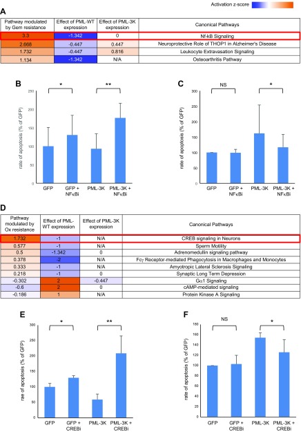
The dismal prognosis of pancreatic ductal adenocarcinoma (PDAC) is mainly due to its rapidly acquired resistance to all conventional treatments. Despite drug-specific mechanisms of resistance, none explains how these cells resist the stress induced by any kind of anticancer treatment. Activation of stress-response pathways relies on the post-translational modifications (PTMs) of involved proteins. Among all PTMs, those mediated by the ubiquitin family of proteins play a central role. Our aim was to identify alterations of ubiquitination, neddylation, and sumoylation associated with the multiresistant phenotype and demonstrate their implications in the survival of PDAC cells undergoing treatment. This approach pointed at an alteration of promyelocytic leukemia (PML) protein sumoylation associated with both gemcitabine and oxaliplatin resistance. We could show that this alteration of PML sumoylation is part of a general mechanism of drug resistance, which in addition involves the abnormal activation of NF-κB and cAMP response element binding pathways. Importantly, using patient-derived tumors and cell lines, we identified a correlation between the levels of PML expression and sumoylation and the sensitivity of tumors to anticancer treatments.-Swayden, M., Alzeeb, G., Masoud, R., Berthois, Y., Audebert, S., Camoin, L., Hannouche, L., Vachon, H., Gayet, O., Bigonnet, M., Roques, J., Silvy, F., Carrier, A., Dusetti, N., Iovanna, J. L., Soubeyran, P. PML hyposumoylation is responsible for the resistance of pancreatic cancer.

Keywords: chemoresistance; post-translational modification; promyelocytic leukemia protein; sumoylation; ubiquitin family.


Conflict of interest statement

The authors thank Dr. Arkaitz Carracedo (Center for Cooperative Research in Bioscience, Spain) for providing PML constructs and to Dr. Guillaume Bossis (IGMM, Montpellier, France) for providing anti-SUMO1 and anti-SUMO2/3 hybridomas. High-throughput sequencing was performed at the Transcriptomique and Génomique Marseille Luminy (TGML) Platform and supported by grants from INSERM, GIS Infrastructures en Biologie Santé et Agronomie (IBiSA), Aix-Marseille Université, and ANR-10-INBS-0009-10. This work was supported by La Ligue Contre le Cancer, Association pour la Recherche sur le Cancer (ARC), Institute National du Cancer (INCa), Cancéropôle Provence-Alpes-Côte d'Azur (PACA), and INSERM. Proteomic analyses were done using the mass spectrometry facility of Marseille Proteomics (marseille-proteomique.univ-amu.fr) and were supported by IBiSA, Plateforme Technologique Aix-Marseille, Cancéropôle PACA, the Institut Paoli-Calmettes, and the Centre de Recherche en Cancérologie de Marseille. The authors declare no conflicts of interest.

Figures

Figure 1
Figure 1
Gemcitabine and oxaliplatin resistance induced alterations of PTM profiles. A) Venn diagram showing the proportions of proteins with gemcitabine resistance–induced alteration of PTMs by 1, 2, or all 3 modifiers (total 186). B) Physical and functional interaction network of proteins with gemcitabine resistance–induced alteration of PTMs. A total of 59 of 186 proteins were found to interact with at least another 1. C) Venn diagram showing the proportions of proteins with oxaliplatin resistance–induced alteration of PTMs by 1, 2, or the 3 modifiers (total 241). D) Physical and functional interaction network of proteins with oxaliplatin resistance–induced alteration of PTMs. A total of 77 out of 241 proteins were found to interact with at least another 1.
Figure 2
Figure 2
Cancer signaling networks in which resistance-induced alterations of ubiquitylation (A), neddylation (B), and sumoylation (C) are involved (according to the Atlas of Cancer Signalling Networks). BER, base excision repair; DR, DNA repair; ECM, extra-cellular matrix; EMT, endothelial mesechymal transition; HR, homologous recombination; MITOCH, mitochondria; MOMP, mitochondrial outer membrane permeabilization; NER, nucleotide excision repair; NHEJ, nonhomologous end joining; TLS, translesion bypass.
Figure 3
Figure 3
Identification of impaired PML sumoylation as a general mechanism of resistance. A) Left panel: Lysates from control and R-Gem cells were subjected to immunoprecipitation using anti-Flag antibody to isolate sumoylated proteins and membranes blotted using an anti-PML antibody to reveal the sumoylated form of PML. Right panel: as in left panel but using lysates from R-Ox cells. B) PML sumoylation kinetics in MiaPaCa-2 cells treated with either gemcitabine or oxaliplatin. After purification of sumoylated protein through immunoprecipitation using anti-Flag antibody, the amount of sumoylated PML was revealed by immunoblotting for PML. C) The previous process as in B but with R-Gem cells. D) The previous process as in B with R-Ox cells. CL, cell lysate; Ctrl, control; IP, immunoprecipitation; NT, non-treated.
Figure 4
Figure 4
PML-repressed sumoylation in resistant cells alters NB formation. A) Representative pictures of immunofluorescence staining of the different MiaPaCa-2 cells in which PML-NBs appear as red spots within nuclei stained with DAPI. B) Quantification and comparison of the number of NBs per nucleus in the MiaPaCa-2 control cells (Ctrl) vs. R-Gem cells and R-Ox cells. C) Quantification and comparison of the mean intensity of NBs in the MiaPaCa-2 parental cells vs. MiaPaCa-2 R-Ox cells and MiaPaCa-2 R-Gem cells. D) Variation of NB intensity in control MiaPaCa-2 cells as well as in R-Gem and R-Ox cells after 24 h of treatment with 10 µM of each drug. *P < 0.05.
Figure 5
Figure 5
PML-repressed sumoylation is involved in chemoresistance of pancreatic cancer cells. A, B) Control MiaPaCa-2 cells were transfected with plasmids expressing WT PML or sumoylation-deficient mutant PML in which the 3 sumoylated lysine residues were changed to arginine or a GFP-expressing vector as a control. The day after transfection, cells were treated with 10 µM gemcitabine or oxaliplatin for an additional 72 h, and then their viability (A) and caspase-3/7 activity (B) were assessed as described in Material and Methods. C, D) MiaPaca-2 R-Gem cells or R-Ox cells were transfected as previously described and were treated with 100 µM gemcitabine or oxaliplatin. After 72 h of treatment, the cell viability (C) and caspase 3/7 activity (D) was assessed as previously described (A, B). Expression of transfected PML was controlled by Western blot (Supplemental Fig. S4).*P < 0.05, **P < 0.02, ***P < 0.01 (Student’s t test analysis from ≥3 independent experiments).
Figure 6
Figure 6
Pathways altered by gemcitabine and oxaliplatin resistances corrected by normal PML sumoylation. RNA-seq data from MiaPaCa-2 control and resistant cells expressing either GFP or PML-WT or PML-3K mutant were analyzed with the help of IPA to identify pathways altered by resistance acquisition to either gemcitabine (A) or oxaliplatin (D) and which is reversed by PML-WT but not by PML-3K expression. A) Table of the main pathways altered by gemcitabine resistance acquisition with the corresponding activation z scores (+ for activation, - for inhibition). B) Control cells were transfected with GFP or PML-3K expression plasmids and treated or not with 5 µM BMS-345541, an NF-κB inhibitor (NF-κBi). C) R-Gem cells transfected with either GFP or PML-WT expression plasmids were treated with gemcitabine 100 ± 1 µM of SRI-22782, an NF-κB activator (NF-κBa). D) Like in A, table of the main pathways altered by oxaliplatin resistance acquisition with the corresponding z scores. E) Control cells were transfected with GFP or PML-3K expression plasmids and treated or not with 0.5 µM of 666-15, a CREB pathway inhibitor (CREBi). F) R-Ox cells transfected with either GFP or PML-WT expression plasmids were treated with oxaliplatin 100 ± 10 µM of Forskolin, a CREB pathway activator (CREBa). For each graph, the rate of apoptosis was quantified using caspase 3/7 activity over the cell viability. NS, no statistical significance. Results are expressed as percentile of GFP nontreated cells, and bars show means ± sd. *P < 0.05, **P < 0.01 (Student’s t test).
Figure 7
Figure 7
PML expression and sumoylation correlates with resistance in patients. A) Lysates from PDXs were subjected to immunoprecipitation using a combination of anti-SUMO1 and anti-SUMO2/3 antibodies followed by PML Western blot analysis. Expression level of PML has also been evaluated by Western blot in lysates. Black arrow: PML; white arrow: sumoylated PML. B) Signals from 2 independent experiments were quantified by densitometry using ImageJ, and PML values were temporized by SUMO values in IP (relative amount of sumoylated PML) or by actin values in lysates (total amount of PML). These values were used to evaluate the correlation between PML sumoylation and expression with patients’ survival. C) The accuracy of PML expression and sumoylation to distinguish patients’ survival rates more or less than 24 mo was tested by an ROC curve (left) and a dot diagram (right). D) Correlation of PML in secretomes of PDX-derived cell lines with their resistant phenotype. The secretion profiles (secretome) as well as the resistant profiles to 6 different chemotherapeutic drugs of a large number of PDX-derived cell lines (chemograms) have been established [(25) and unpublished results]. For each considered drug, the amount of secreted PML protein in the 10 most sensitive cell lines and the 10 most resistant cell lines was quantified by mass spectrometry. 5FU, 5 Fluorouracil; AUC, area under the curve; IP, immunoprecipitation; Sens, sensitivity; SN38, 7-ethyl-10-hydroxycamptothecin; Spec, specificity; THOP1, thimet oligopeptidase 1; TXT, docetaxel. Values are presented in box plots with result from Student’s t test analysis.

Similar articles

Cited by

References

    1. Schneider G., Siveke J. T., Eckel F., Schmid R. M. (2005) Pancreatic cancer: basic and clinical aspects. Gastroenterology 128, 1606–1625 - PubMed
    1. Siegel R. L., Miller K. D., Jemal A. (2018) Cancer statistics, 2018. CA Cancer J. Clin. 68, 7–30 - PubMed
    1. Conroy T., Desseigne F., Ychou M., Bouché O., Guimbaud R., Bécouarn Y., Adenis A., Raoul J. L., Gourgou-Bourgade S., de la Fouchardière C., Bennouna J., Bachet J. B., Khemissa-Akouz F., Péré-Vergé D., Delbaldo C., Assenat E., Chauffert B., Michel P., Montoto-Grillot C., Ducreux M.; Groupe Tumeurs Digestives of UnicancerPRODIGE Intergroup (2011) FOLFIRINOX versus gemcitabine for metastatic pancreatic cancer. N. Engl. J. Med. 364, 1817–1825 - PubMed
    1. Von Hoff D. D., Ervin T., Arena F. P., Chiorean E. G., Infante J., Moore M., Seay T., Tjulandin S. A., Ma W. W., Saleh M. N., Harris M., Reni M., Dowden S., Laheru D., Bahary N., Ramanathan R. K., Tabernero J., Hidalgo M., Goldstein D., Van Cutsem E., Wei X., Iglesias J., Renschler M. F. (2013) Increased survival in pancreatic cancer with nab-paclitaxel plus gemcitabine. N. Engl. J. Med. 369, 1691–1703 - PMC - PubMed
    1. Adamska A., Elaskalani O., Emmanouilidi A., Kim M., Abdol Razak N. B., Metharom P., Falasca M. (2018) Molecular and cellular mechanisms of chemoresistance in pancreatic cancer. Adv. Biol. Regul. 68, 77–87 - PubMed

Publication types

LinkOut - more resources