Review
. 2019 Sep 25;103(6):980-1004.
doi: 10.1016/j.neuron.2019.07.009.

Development and Arealization of the Cerebral Cortex

Affiliations
Review

Development and Arealization of the Cerebral Cortex

Cathryn R Cadwell et al. Neuron. .

Abstract

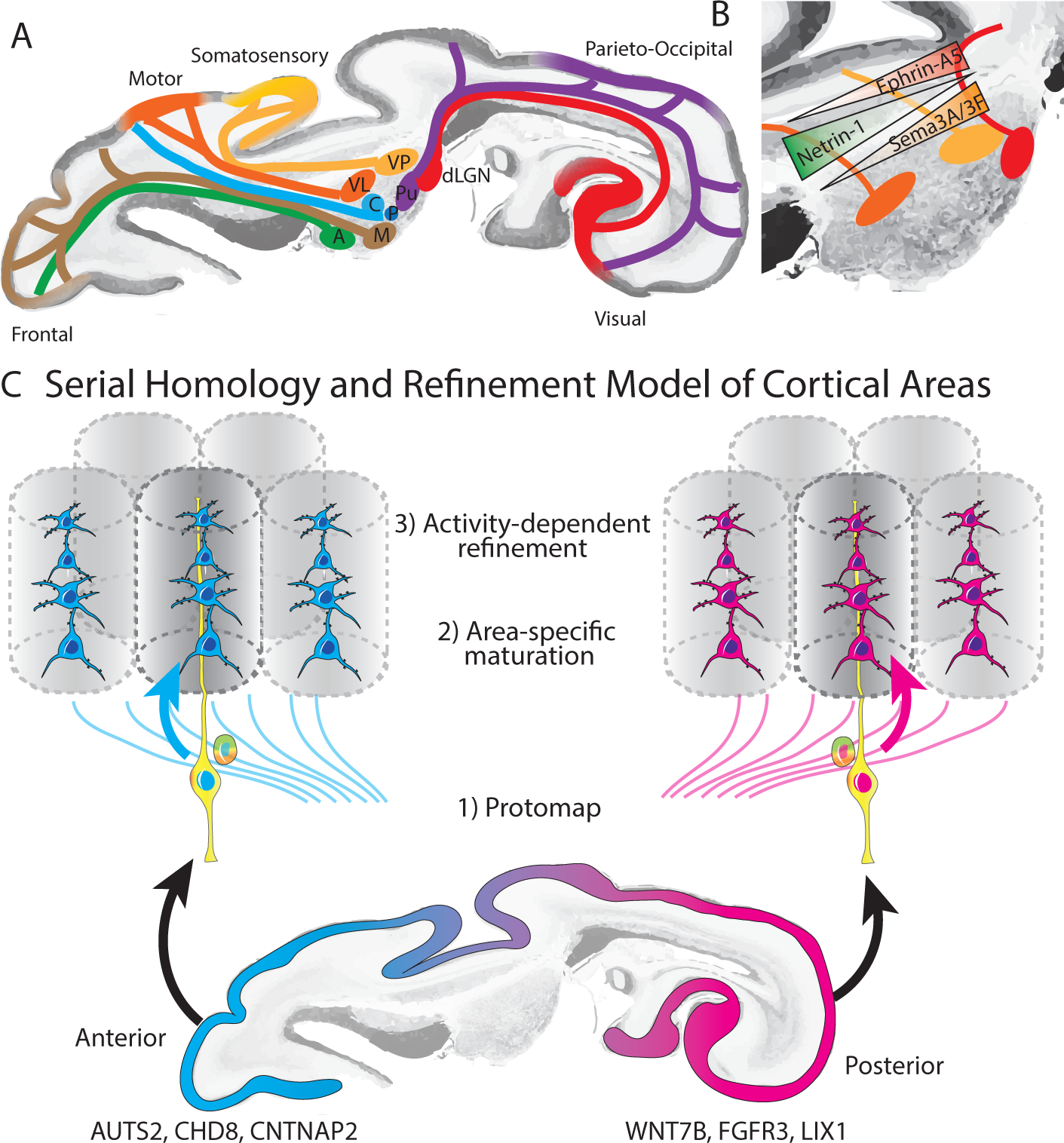
Adult cortical areas consist of specialized cell types and circuits that support unique higher-order cognitive functions. How this regional diversity develops from an initially uniform neuroepithelium has been the subject of decades of seminal research, and emerging technologies, including single-cell transcriptomics, provide a new perspective on area-specific molecular diversity. Here, we review the early developmental processes that underlie cortical arealization, including both cortex intrinsic and extrinsic mechanisms as embodied by the protomap and protocortex hypotheses, respectively. We propose an integrated model of serial homology whereby intrinsic genetic programs and local factors establish early transcriptomic differences between excitatory neurons destined to give rise to broad "proto-regions," and activity-dependent mechanisms lead to progressive refinement and formation of sharp boundaries between functional areas. Finally, we explore the potential of these basic developmental processes to inform our understanding of the emergence of functional neural networks and circuit abnormalities in neurodevelopmental disorders.

Keywords: autism; brain development; cerebral cortex; human brain; machine learning; neural networks; neurogenesis; protocortex; protomap; serial homology.


Conflict of interest statement

Declaration of Interests

The authors declare no competing interests.

Figures

Figure 1:
Figure 1:. Arealization of the human cerebral cortex.
A) Classical cytoarchitectonic areas described by (Brodmann, 1909). B) Areal differences in local microcircuit architecture between granular and agranular cortices modified from (Beul and Hilgetag, 2015; Shipp, 2005). C) Hierarchical organization between cortical areas, inferred and/or modified from (Badre and Nee, 2018; Felleman and Van Essen, 1991; Ventre-Dominey, 2014).
Figure 2:
Figure 2:. Timing of neurodevelopmental events in the cerebral cortex.
A) Key patterning centers set up morphogen and transcription factor gradients across the developing neuroepithelium to influence area-specific cell fates. B) Schematic comparison of the mouse and human cerebral cortical development at peak stages of neurogenesis. While the processes are broadly conserved, several important differences can be highlighted: prominent expansion of the outer subventricular zone (OSVZ) and of the outer radial glia population which resides therein (Fietz et al., 2010; Hansen et al., 2010; Reillo et al., 2011); expansion of the subplate (SP), which is associated with the expansion of the axonal plexus, but not necessarily cell numbers (Duque et al., 2016); expansion of the upper layer excitatory neuron types (Hodge et al., 2018); discontinuation of the radial glia scaffold (Nowakowski et al., 2016); emergence of persisting Cajal-Retzius cells (Meyer and Gonzalez-Gomez, 2018); expansion of axonal plexus in the OSVZ and the emergence of multilaminar axonal-cellular compartment (Zunic Isasegi et al., 2018); early arrival of thalamocortical plexus in the cortical anlage (Marin-Padilla, 1983). C) Broad comparison of key neurogenesis periods across species, including data from (Clancy et al., 2001; Rakic, 1974). Embryonic days (E) next to species names indicate approximate length of gestation.
Figure 3:
Figure 3:. Maturation and differentiation in the cortex.
A) Left: schematic representing radial glia maturation from neuroepithelial stem cells (NESC), followed by their differentiation into astrocytes. Right: schematic representing sequential production of cortical layers from radial glia in the mouse. B) in contrast, human cortical development involves an expanded diversity of radial glia with distinct maturation trajectories (left). Right: neurogenesis in the human cortex occurs in the ventricular zone early in development, and progressively shifts towards the outer subventricular zone. VZ-ventricular zone, SVZ - subventricular zone, ISVZ- inner subventricular zone, OSVZ- outer subventricular zone, tRG-truncated radial glia, vRG- ventricular radial glia, NESC- neuroepithelial stem cells, IPC- intermediate progenitor cells, oRG- outer radial glia, WM- white matter, LI-VI - cortical layer I-VI.
Figure 4:
Figure 4:. Mechanisms of arealization.
A) Early development of thalamocortical tracts provides anatomical basis for modality specific responses in the cortex, activity dependent changes in cortical area size and functional circuit development. B) Morphogen gradients contribute to shaping thalamic areal specification. C) Serial homology and refinement model, in which area-specific gene expression programs establish an initial “protomap” which is further refined by area-specific maturation signals and activity-dependent processes to generate the final mature cortical areas.

References

    1. Adesnik H, and Naka A (2018). Cracking the Function of Layers in the Sensory Cortex. Neuron 100, 1028–1043. - PMC - PubMed
    1. Adler A, Zhao R, Shin ME, Yasuda R, and Gan W-B (2019). Somatostatin-Expressing Interneurons Enable and Maintain Learning-Dependent Sequential Activation of Pyramidal Neurons. Neuron 102, 202–216.e7. - PMC - PubMed
    1. Aevermann BD, Novotny M, Bakken T, Miller JA, Diehl AD, Osumi-Sutherland D, Lasken RS, Lein ES, and Scheuermann RH (2018). Cell type discovery using single-cell transcriptomics: implications for ontological representation. Hum. Mol. Genet 27, R40–R47. - PMC - PubMed
    1. Alcamo EA, Chirivella L, Dautzenberg M, Dobreva G, Farinas I, Grosschedl R, and McConnell SK (2008). Satb2 regulates callosal projection neuron identity in the developing cerebral cortex. Neuron 57, 364–377. - PubMed
    1. Alzu’bi A, Homman-Ludiye J, Bourne JA, and Clowry GJ (2019). Thalamocortical Afferents Innervate the Cortical Subplate much Earlier in Development in Primate than in Rodent. Cereb. Cortex 29, 1706–1718. - PMC - PubMed

Publication types