Review
. 2019 Nov;63(4):R93-R102.
doi: 10.1530/JME-19-0153.

Circadian clock genes and the transcriptional architecture of the clock mechanism

Affiliations
Review

Circadian clock genes and the transcriptional architecture of the clock mechanism

Kimberly H Cox et al. J Mol Endocrinol. 2019 Nov.

Abstract

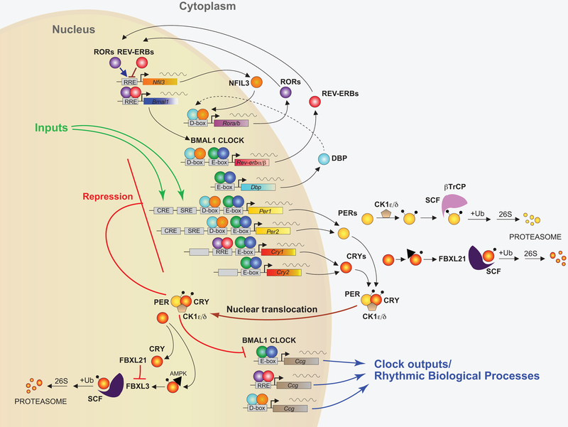
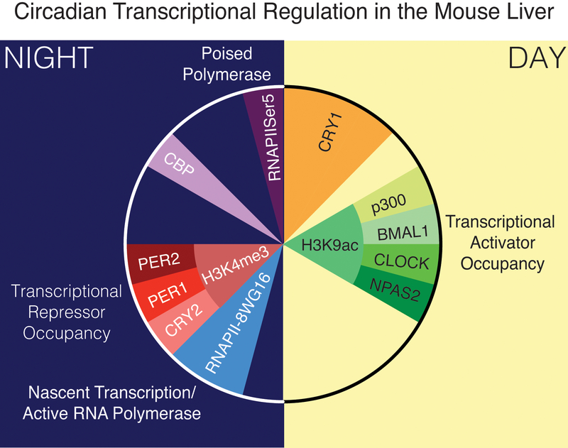
The mammalian circadian clock has evolved as an adaptation to the 24-h light/darkness cycle on earth. Maintaining cellular activities in synchrony with the activities of the organism (such as eating and sleeping) helps different tissue and organ systems coordinate and optimize their performance. The full extent of the mechanisms by which cells maintain the clock are still under investigation, but involve a core set of clock genes that regulate large networks of gene transcription both by direct transcriptional activation/repression as well as the recruitment of proteins that modify chromatin states more broadly.

Keywords: circadian clock; clock genes; mammalian; transcription.


Figures

Figure 1:
Figure 1:. Core components of the mammalian circadian clock.
In the core feedback loop, the transcription factors BMAL1 (green circles) and CLOCK (blue circles) bind to E-box domains on gene promoters, including the genes for Per1 and Per2 (yellow) and Cry1 and Cry2 (red/yellow). PERs (yellow circles) and CRYs (red/yellow circles) dimerize and translocate to the nucleus after binding with casein kinase δ (CK1δ) or CK1ϵ, where they repress their own transcription. The stability of PER and CRY is regulated both in the cytoplasm and within the nucleus by several proteins, including FBXL21 and FBXL3. In a second feedback loop, CLOCK and BMAL1 also regulate the transcription of genes for the nuclear receptors REV-ERBα and REV-ERBβ (red circles), which compete with the retinoic acid-related orphan receptors, RORα, RORβ, and RORγ (purple circles) for binding to RRE elements on the BMAL1 gene promoter, providing both positive (ROR) and negative (REV-ERB) regulation of BMAL1 transcription. A third feedback loop is mediated by CLOCK/BMAL1-mediated transcription of the gene Dbp (light blue) and the ROR/REV-ERB-mediated transcription of Nfil3 (orange). DBP (light blue circles) and NFIL3 (orange circles) dimerize and bind to D-box elements on the promoters of many of the core clock genes, providing additional layers of regulation. In addition, CLOCK/BMAL1, ROR/REV-ERB, and DBP/NFIL3 regulate the transcription of many other clock output genes (Figure modified from (Takahashi, 2017)).
Figure 2:
Figure 2:. 24-hour depiction of genome-wide circadian transcriptional regulation in the mouse liver.
Peak occupancy of transcriptional activators at gene promoters occurs in the middle of the day and corresponds with a peak in H3K9acetylation. Peak transcription occurs shortly after nightfall, as indicated by activated RNA polymerase binding. Transcriptional repressor occupancy peaks shortly thereafter, and corresponds with a peak in Hk34 tri-methylation. Additional transcription factors and co-factors, such as CRY1 and CBP appear to occupy promoters at different times, and poised RNA polymerase occupancy peaks just at the end of the 24-hour cycle (Based on data from (Koike et al., 2012) and figure from (Takahashi, 2017)).
Figure 3:
Figure 3:. BMAL1 regulation of metabolism.
Overlay of BMAL1 target genes (indicated in red) on diverse metabolic pathways in the liver. BMAL1 occupancy data are from a previously published ChIP-seq dataset (Koike et al., 2012). The original metabolic pathway is from a KEGG analysis (used with permission) and has been simplified to show major nodes (Kanehisa and Goto, 2000, Kanehisa et al., 2017). In KEGG, nodes indicate enzymes and lines indicate connections in metabolic pathways, with colors indicating pathways serving similar functions. The red dots and lines indicate BMAL1 interactions with genes involved in these pathways.

References

    1. ABRAHAMSON EE & MOORE RY 2001. Suprachiasmatic nucleus in the mouse: retinal innervation, intrinsic organization and efferent projections. Brain Res, 916, 172–91. - PubMed
    1. ABRUZZI KC, RODRIGUEZ J, MENET JS, DESROCHERS J, ZADINA A, LUO W, TKACHEV S & ROSBASH M 2011. Drosophila CLOCK target gene characterization: implications for circadian tissue-specific gene expression. Genes Dev, 25, 2374–86. - PMC - PubMed
    1. ACOSTA-RODRIGUEZ VA, DE GROOT MHM, RIJO-FERREIRA F, GREEN CB & TAKAHASHI JS 2017. Mice under Caloric Restriction Self-Impose a Temporal Restriction of Food Intake as Revealed by an Automated Feeder System. Cell Metab, 26, 267–277 e2. - PMC - PubMed
    1. ALBRECHT U 2012. Timing to perfection: the biology of central and peripheral circadian clocks. Neuron, 74, 246–60. - PubMed
    1. ASHER G, GATFIELD D, STRATMANN M, REINKE H, DIBNER C, KREPPEL F, MOSTOSLAVSKY R, ALT FW & SCHIBLER U 2008. SIRT1 regulates circadian clock gene expression through PER2 deacetylation. Cell, 134, 317–28. - PubMed

Publication types